Submandibular fossa (Latin: fovea submandibularis)
Also called the submandibular fovea
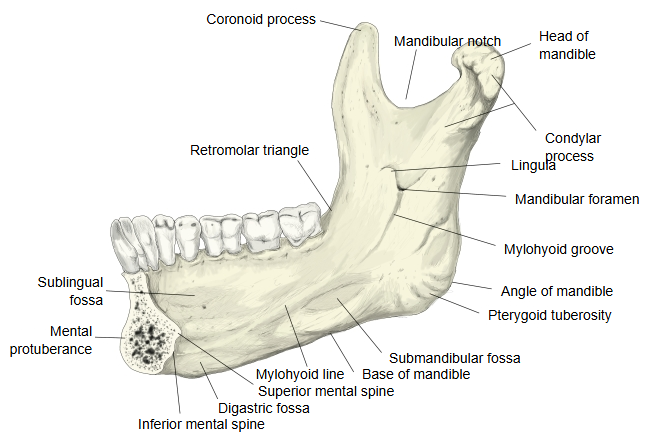
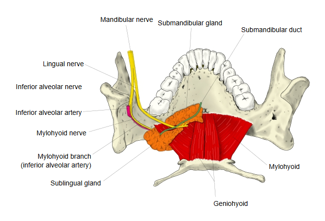
It lies on the inner surface of the body of the mandible, between the mylohyoid line above and the base of the mandible below, roughly beneath the molar teeth. In front of it, above the mylohyoid line, is the sublingual fossa for the sublingual gland; the two fossae are separated by the line and by the mylohyoid attached to it. The part of the submandibular gland that lies below the mylohyoid (its superficial part) fits into the fossa. Above and behind it the mylohyoid groove runs down from the mandibular foramen. The depth of the fossa varies a good deal between individuals.
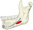 Submandibular fossa (medial view)
 Mandible (medial surface)
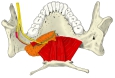 Floor of mouth (superior view)
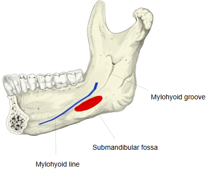
Reference websites
[Wikipedia]
· Submandibular fovea, Wikipedia.
· Mandible, Wikipedia.
[Websites with illustrations]
· Mandible, inner surface, side view (Gray's Anatomy, fig. 177) (Wikimedia Commons)

· The mandible: structure and attachments (TeachMeAnatomy)
· The mandible: anatomy, structure, function (Kenhub)
Related terms
References and sources
- Further reading: Submandibular fovea, Wikipedia.
- Further reading: Mandible, Wikipedia.
- Federative International Programme for Anatomical Terminology (FIPAT). Terminologia Anatomica, 2nd ed. (TA2), 2019.