Trochlea of superior oblique (Latin: trochlea musculi obliqui superioris)
Also called: trochlea of orbit
A small pulley through which the tendon of the superior oblique passes and is held in place. It is attached to the trochlear fovea, a small pit at the front medial corner of the orbital surface of the orbital part of the frontal bone, just behind the upper medial margin of the orbit; sometimes a small trochlear spine is present instead.
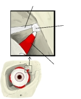 Trochlea (anterior view)
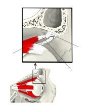 Trochlea (lateral view)
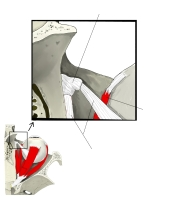 Trochlea (superior view)
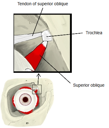
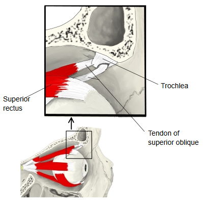
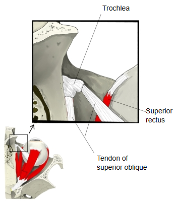
Few textbooks describe it in detail. It is a ring of fibrous tissue with cartilage in its inner part, and a synovial sheath lines the tendon where it slides through. The superior oblique runs forwards along the upper medial wall of the orbit; its tendon passes through the trochlea, turns sharply backwards and laterally, and inserts on the eyeball behind its equator, under the superior rectus. Because of this change in direction the muscle depresses, abducts and medially rotates (intorts) the eye. Its nerve, the trochlear nerve, is named after the trochlea.
Related terms
References and sources
- Further reading: Trochlea of superior oblique, Wikipedia.
- Further reading: Superior oblique muscle, Wikipedia.
- Federative International Programme for Anatomical Terminology (FIPAT). Terminologia Anatomica, 2nd ed. (TA2), 2019.