Endolymphatic duct (Latin: ductus endolymphaticus)
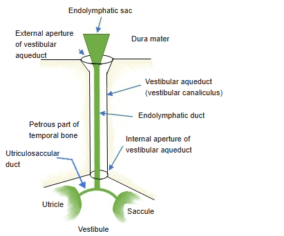
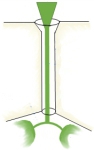 Utriculosaccular duct to endolymphatic sac (schematic)
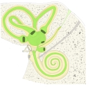 Bony and membranous labyrinth
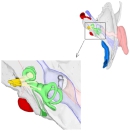 Right inner ear (from above)
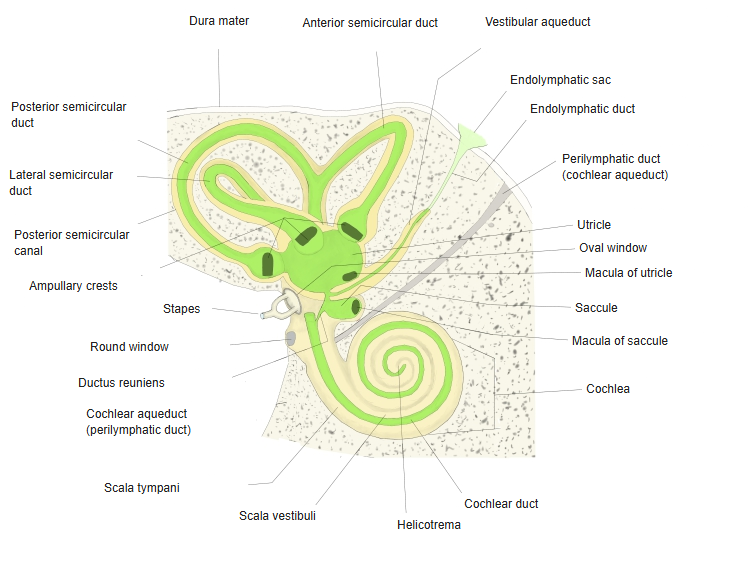
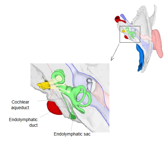
Rauber-Kopsch's anatomy describes it as follows (summary):
The walls of the utricle and the saccule touch at one place, but they do not fuse into a single partition; they stay separate. The two sacs are joined by one long, thin duct, open at both ends, with an internal diameter of 0.17 mm: the endolymphatic duct (ductus endolymphaticus). Its limb to the saccule is the thicker one; the thinner limb, which opens into the utricle, is the utriculosaccular duct. At its end the endolymphatic duct widens into a fairly large, flat pouch, the endolymphatic sac, about 1 cm long and 5–8 mm wide. The sac lies beyond the opening of the vestibular aqueduct (canaliculus vestibuli), against the posterior surface of the petrous part of the temporal bone, between two layers of the dura mater, and points downward and laterally.
The endolymphatic duct and sac belong to the endolymphatic space of the internal ear (compare the perilymphatic duct of the perilymphatic space). Disorders of the duct and sac are linked with Ménière's disease and with an enlarged vestibular aqueduct.
Reference websites
[Wikipedia]
· Endolymphatic duct, Wikipedia.
· Vestibular aqueduct, Wikipedia.
[Websites with illustrations]
· Endolymphatic duct in the aqueduct of the vestibule (schematic) (Pinterest)

· Endolymphatic duct and sac, vestibular aqueduct (schematic) (X (Twitter))

Related terms
- Endolymphatic sac
- Endolymphatic space
- Membranous labyrinth
- Saccule
- Utricle
- Vestibule
- Perilymphatic duct
- Petrous part
- Internal ear
References and sources
- Further reading: Endolymphatic duct, Wikipedia.
- Further reading: Vestibular aqueduct, Wikipedia.
- Federative International Programme for Anatomical Terminology (FIPAT). Terminologia Anatomica, 2nd ed. (TA2), 2019.