Pia mater (Latin: pia mater)
Also called Pia
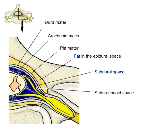
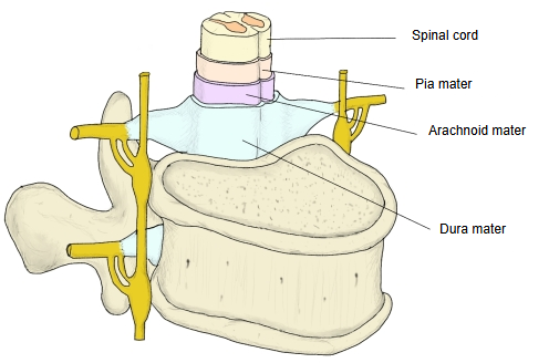
- Dura mater : the outermost and toughest layer.
- Arachnoid mater : the layer between the dura and the pia; it has no blood vessels of its own.
- Pia mater : the innermost, very thin layer, made of an inner and an outer layer.
- Epidural (extradural) space : outside the dura: in the vertebral canal, the space between the spinal dura and the wall of the canal.
- Dura mater
- Subdural space : between the dura and the arachnoid; it appears only in disease, for example after an injury.
- Arachnoid mater
- Subarachnoid space : between the arachnoid and the pia; filled with cerebrospinal fluid.
- Pia mater
Cranial pia mater
· The cranial pia mater covers the brain and dips into its sulci and fissures. It also reaches into the ventricles, the spaces inside the brain: in the roof of the lateral, third and fourth ventricles, pia and the lining of ependymal cells are joined together and form the tela choroidea of each ventricle.
· The tela choroidea carries the choroid plexuses, which are very rich in blood vessels; they secrete most of the cerebrospinal fluid. The tela choroidea also holds the choroid plexuses, which hang into the ventricles, in place.
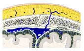 Meninges of the brain
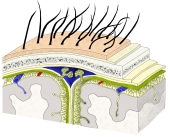 Layers from scalp to brain
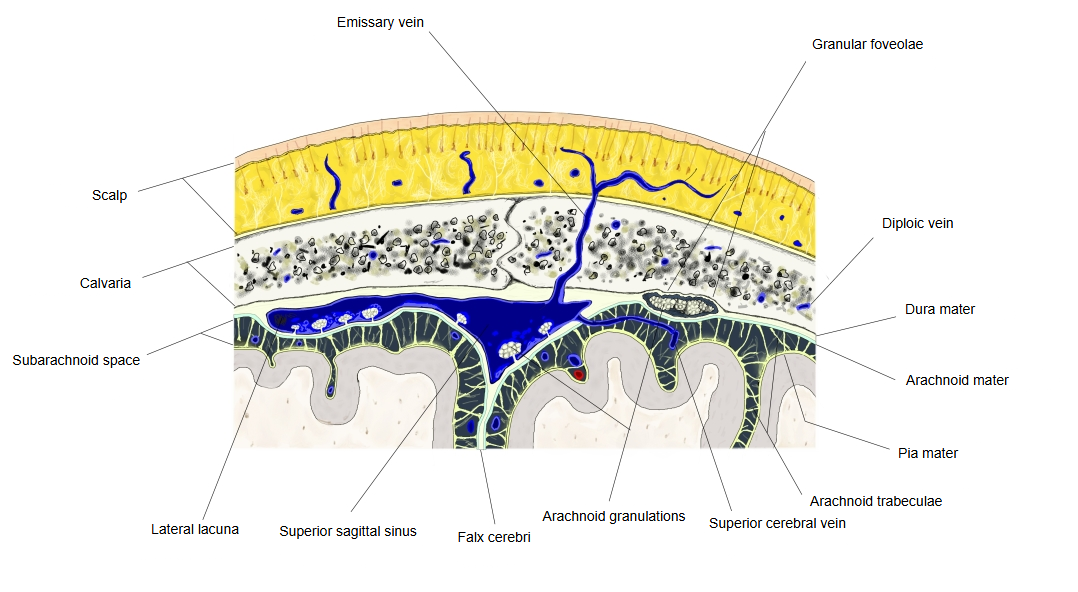
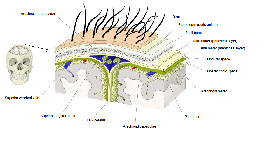
· Blood vessels run on the pia in the subarachnoid space. Where they enter the brain, the pia follows them a short way, so a narrow perivascular space lies around each vessel.
Spinal pia mater
· The spinal pia mater is thicker and firmer than the cranial pia. It covers the whole surface of the spinal cord and enters every groove, including the anterior median fissure. It is divided into the intima pia and the epipial layer, and the arteries and veins of the spinal cord run between these two layers.
· Below the end of the spinal cord the pia continues downward as a thin thread, the filum terminale. Its pial part (filum terminale internum) runs to the lower end of the dural sac; there it takes on a covering of dura and continues as the dural part (filum terminale externum), which is anchored to the back of the coccyx.
· On each side of the spinal cord the pia forms the saw-toothed denticulate ligament. Its teeth pass between the anterior and posterior roots and attach through the arachnoid to the dura, holding the cord in place. There are about 20 pairs along the length of the cord.
 Meninges of the spinal cord
 Spinal cord and its coverings
Epipial layer
· The epipial layer is the outer layer of the pia: a network of bundles of collagen fibres that is continuous with the trabeculae of the arachnoid mater outside it.
· It is well developed on the spinal cord, where the vessels of the cord run within it, but it is largely missing over the gyri of the cerebral cortex. There the vessels that supply the brain run on the surface of the intima pia, held in place by the arachnoid trabeculae.
Intima pia
· The intima pia is the inner layer: a thin, continuous sheet of fine reticular fibres and elastic fibres that follows the shape of the brain and spinal cord closely and is firmly attached to their nervous tissue.
· Where blood vessels enter or leave the central nervous system, the intima pia is folded in with them for a short distance, leaving the perivascular space between the vessel and the pia.
· The intima pia has no blood vessels of its own; it is nourished by the cerebrospinal fluid and by the nervous tissue just beneath it.
Reference websites
[Wikipedia]
· Pia mater, Wikipedia.
· Meninges, Wikipedia.
[Websites with illustrations]
· Pia mater on the spinal cord (histology slide) (University of Oklahoma Health Sciences Center)

Related terms
- Cerebral ventricles
- Rhomboid fossa
- Arachnoid mater
- Dura mater
- Subarachnoid space
- Choroid plexus
- Pial part of filum terminale
- Denticulate ligament
- Spinal cord