Middle cribriform area (Latin: macula cribrosa media)
The middle cribriform area is one of the maculae cribrosae on the medial wall of the vestibule; it is found in the spherical recess.
Maculae cribrosae: groups of small holes that let through the nerve fibres coming from the vestibular ganglionSpherical recess: the hollow that holds the saccule
Holes in the
medial wall of the
vestibule that
let nerves through
medial wall of the
vestibule that
let nerves through
- Superior cribriform area : Site: above the pyramid of the vestibule and in the adjacent elliptical recess. Nerve: the utriculo-ampullary nerve, which it leads to the superior vestibular area.
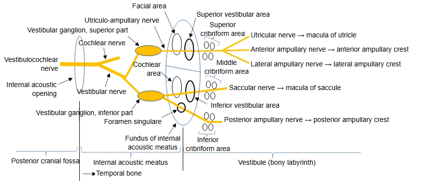
- Middle cribriform area : Site: the floor of the spherical recess. Nerve: the saccular nerve, which it leads to the inferior vestibular area.
- Inferior cribriform area : Site: from near the cochlear recess to the opening of the bony ampulla of the posterior semicircular canal. Nerve: the posterior ampullary nerve, which it leads to the foramen singulare.
Through the middle cribriform area passes the saccular nerve, which comes through the inferior vestibular area of the fundus of the internal acoustic meatus.
It comes from the lower part of the vestibular ganglion; after passing through, it is distributed to the sensory epithelium of the macula of the saccule
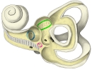 Maculae cribrosae
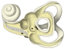 Left bony labyrinth, inner surface (opened from the lateral side)
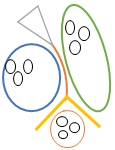 Maculae cribrosae, recesses and the vestibular crest (model)
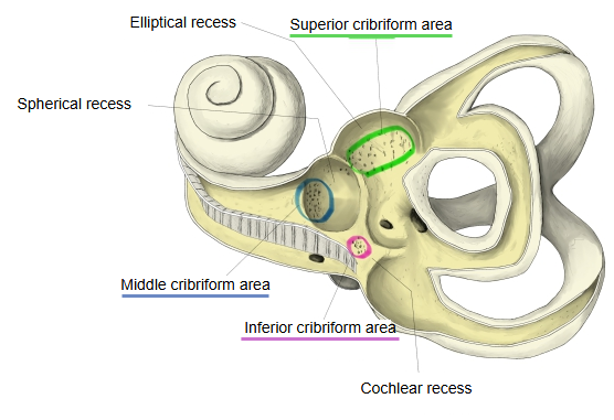
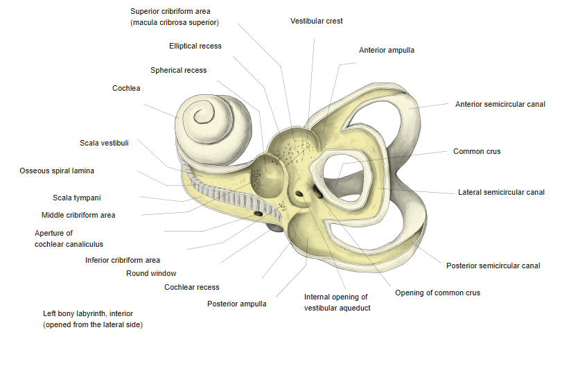
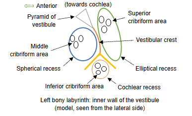
The part of the floor of the spherical recess that lets the saccular nerve through is called the middle cribriform area. It lies at the front and lower part of the recess, and on the other side of this thin wall it opens into the inferior vestibular area of the internal acoustic meatus.
Reference websites
[Wikipedia]
· Bony labyrinth, Wikipedia.
· Vestibule of the ear, Wikipedia.
Related terms
- Spherical recess
- Superior cribriform area
- Inferior cribriform area
- Macula of saccule
- Saccular nerve
- Vestibule
- Vestibular ganglion
References and sources
- Further reading: Bony labyrinth, Wikipedia.
- Federative International Programme for Anatomical Terminology (FIPAT). Terminologia Anatomica, 2nd ed. (TA2), 2019.