Intervertebral foramen (Latin: foramen intervertebrale)
Also called the neural foramen
- Overview
- Number and details
- Reference websites
- (Wikipedia, websites with illustrations)
- Related terms
Overview
· Although it is called a foramen (a hole), it is a short canal with some depth: a three-dimensional space rather than a simple hole.
Its walls are: above and below, the pedicles of the two vertebrae (their vertebral notches); in front, the backs of the two vertebral bodies and the intervertebral disc between them; behind, the zygapophysial (facet) joint between the articular processes. Outside the foramen, on the cervical vertebrae, the nerve runs on in the groove for the spinal nerve on the transverse process.
Disc herniation, bony outgrowths or thickened ligaments can narrow the foramen and compress the nerve (foraminal stenosis).
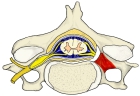 Vertebra and spinal nerve (superior view)
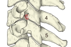 Intervertebral foramen (cervical vertebrae)
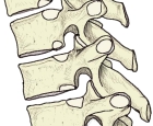 Intervertebral foramina (thoracic vertebrae)
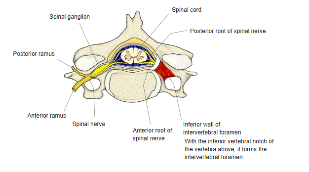
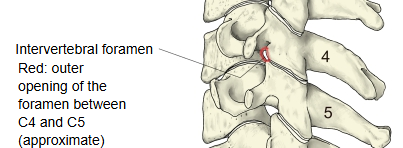
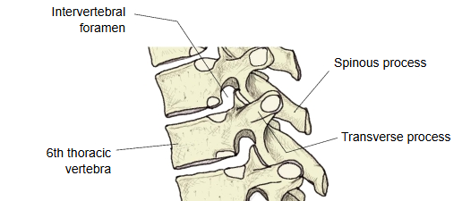
Number and details
The spinal nerves, each with its own spinal ganglion, are counted by region as follows. One pair of intervertebral foramina lies between each two neighbouring cervical, thoracic and lumbar vertebrae; the 1st cervical nerve leaves above the atlas, between it and the occipital bone, the 1st to 4th sacral nerves leave through the four pairs of sacral foramina, and the 5th sacral nerve and the coccygeal nerve through the sacral hiatus.
| Region | Number (left and right pairs) | |
| 1 | Cervical | 8 pairs (C1–C8) |
| 2 | Thoracic | 12 pairs (T1–T12) |
| 3 | Lumbar | 5 pairs (L1–L5) |
| 4 | Sacral | 5 pairs (S1–S5) |
| 5 | Coccygeal | 1 pair (Co1) |
| Total | 31 pairs (62 in all) | |
Size
The table below describes the size of the intervertebral foramina in adults by region (approximate values).
Note: the size varies with build and sex, and also with level, posture and movement (the foramen narrows when the spine is bent backwards).
| Size | Features | ||
| 1 | Cervical | small; at C5–C6 about 8–10 mm high and about 5 mm wide | The nerve root runs obliquely forwards and outwards and fills much of the foramen; the neck moves a lot, so the foramen narrows easily (for example with bony outgrowths). |
| 2 | Thoracic | relatively large compared with the small thoracic nerves | The thoracic spine moves little, so the foramina are stable; the rib heads lie close to them. |
| 3 | Lumbar | the largest; about 18–23 mm high and 8–10 mm wide | Large openings for the thick lumbar nerve roots, which descend in the cauda equina before they leave. |
| 4 | Sacral | the 1st to 4th sacral nerves leave through the sacral foramina; the 5th, with the coccygeal nerve, through the sacral hiatus | In the fused sacrum the intervertebral foramina lie inside the bone and open through the anterior and posterior sacral foramina, a different arrangement from the true intervertebral foramina. |
Contents
The table below lists the structures found in the intervertebral foramen.
| Structure | Description | |
| 1 | Spinal nerve | the mixed nerve formed where the anterior (motor) and posterior (sensory) roots from the spinal cord join |
| 2 | Spinal ganglion | the swelling on the posterior root holding the cell bodies of the sensory neurons (dorsal root ganglion); the largest structure in the foramen, usually lying in it or just inside it |
| 3 | Spinal arteries and intervertebral veins | spinal branches of the segmental arteries supply the vertebral column and its contents with blood, and the intervertebral veins drain it; they often run with the nerve |
| 4 | Connective tissue around the nerve | soft connective tissue that protects and supports the nerves and vessels; the small recurrent meningeal (sinuvertebral) nerve also runs back through the foramen into the vertebral canal |
| 5 | Adipose tissue | a small amount of fat that cushions and protects the contents |
What leaves through it
| Item | Description | |
| 1 | Spinal nerve | the mixed nerve (motor and sensory) formed by the joining of the posterior and anterior roots; it passes through the foramen to the periphery |
| 2 | Spinal ganglion | usually lies at the inner end of the foramen or in it; from it the nerve fibres run outwards (the cell bodies are here) |
| 3 | Branches of the spinal nerve | just after leaving the foramen the spinal nerve divides into the anterior and posterior rami, which go to the front and the back of the body |
| 4 | Rami communicantes of the sympathetic system | the white and grey rami communicantes join the spinal nerve (its anterior ramus) to the sympathetic trunk, just outside the foramen |
| 5 | Branches of the intervertebral vessels | the spinal branches of the segmental arteries enter the vertebral canal through the foramen, and the intervertebral veins leave through it |
Reference websites
[Wikipedia]
· Intervertebral foramen, Wikipedia.
· Dorsal root ganglion, Wikipedia.
[Websites with illustrations]
· Intervertebral foramen: anatomy and function (Kenhub)
· The vertebral column (TeachMeAnatomy)
Related terms
- Intervertebral disc
- Vertebra
- Pedicle of vertebral arch
- Superior vertebral notch
- Inferior vertebral notch
- Spinal nerves
- Spinal ganglion
- Vertebral canal
- Intervertebral veins
- Groove for spinal nerve
References and sources
- Further reading: Intervertebral foramen, Wikipedia.
- Further reading: Dorsal root ganglion, Wikipedia.
- Federative International Programme for Anatomical Terminology (FIPAT). Terminologia Anatomica, 2nd ed. (TA2), 2019.