Mitochondrion (Latin: mitochondrion)
Also called Mitochondria (plural)
One of the structures inside each cell of our body (the organelles): the factory that produces the energy we need for all our activities.
of the
cell
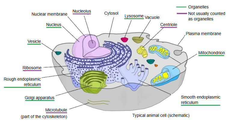
- Cell membrane : phospholipid bilayer, membrane proteins, sugar chains.
- Cytoplasm
- Cytosol : water, electrolytes, metabolic enzymes, free amino acids, metabolic intermediates.
- Organelles : mitochondria, endoplasmic reticulum, centrosome, Golgi apparatus, lysosomes, peroxisomes, vesicles (some sources do not count vesicles as organelles).
- Cytoskeleton : microtubules, actin filaments, intermediate filaments.
- Nucleus : nuclear envelope, nucleoplasm, chromatin, nucleolus; the nucleus is also counted among the organelles.
More details
[About a tenth of body weight in total]
Our body is made of some tens of trillions of cells, and mitochondria are present in almost all of them (mature red blood cells are the main exception). Their total mass is said to be about a tenth of body weight; in liver cells they make up about 22% of the cell.
Shape
[Surprisingly varied shapes?]
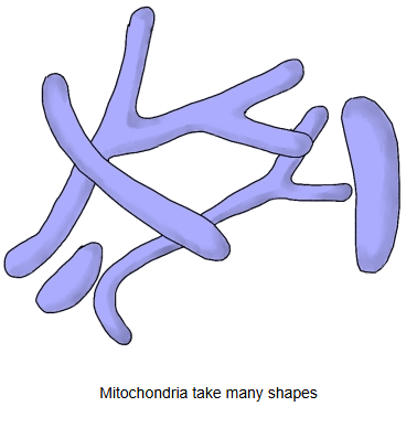
The name comes from Greek: "mito-" means thread and "chondrion" a small grain. Pictures usually show a capsule shape, but mitochondria constantly fuse and divide inside the cell, so their real shape is more complex.
Size and range of shapes:
They are usually about 0.5 µm wide, but their shape varies with the organism and the state of the cell: round, rod-shaped, thread-like, branched or networked, and not rarely up to about 10 µm long.
Number
[100 to 3,000]
In humans one cell has from about 100 to 3,000, a wide range. As they are the energy factories, cells that work hard have more of them, especially cardiac and skeletal muscle fibres, nerve cells and liver cells.
The number also differs greatly between kinds of cell:
A human sperm has only a few dozen mitochondria, packed into its midpiece, whereas an oocyte (egg cell) has around 100,000. The large store in the oocyte is handed on to the embryo, which relies on it during the first cell divisions after fertilization.
Structure and components
A mitochondrion is enclosed by a double membrane. Its main components are listed below; it also contains many other proteins and enzymes.
| Component | Description | ||
|---|---|---|---|
| 1 | Inner membrane | The inner of the two membranes; folded into cristae; carries the electron transport chain and ATP synthase | 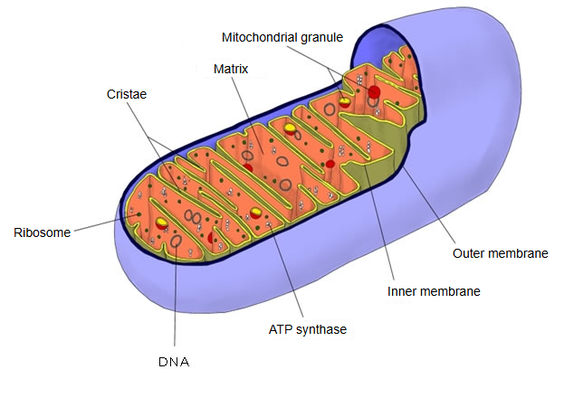 |
| 2 | Outer membrane | The outer membrane: smooth and porous; it encloses the whole organelle | |
| 3 | ATP synthase | An enzyme in the inner membrane that makes ATP | |
| 4 | DNA | Its own circular DNA; hundreds to thousands of copies per cell | |
| 5 | Ribosomes | Factories for proteins (its own ribosomes) | |
| 6 | Cristae | Shelf-like folds of the inner membrane projecting inwards | |
| 7 | Matrix | The space inside the inner membrane | |
| 8 | Mitochondrial granules | Granules in the matrix |
of a
mitochondrion
- Outer membrane : a smooth, porous unit membrane (lipid bilayer).
- Intermembrane space : the narrow space between the outer and inner membranes (hydrogen ions = protons are pumped into it).
- Inner parts
- Inner membrane : a lipid bilayer folded inwards into cristae; it carries the electron transport chain (the energy-producing enzymes) and ATP synthase.
- Matrix
- DNA : the mitochondrion's own circular DNA, which holds genetic information.
- Ribosomes : the mitochondrion's own; they make 13 membrane proteins, which are built into the inner membrane.
- RNA : mainly the three kinds of RNA used in protein synthesis (mRNA, tRNA, rRNA).
- Enzymes and other contents : soluble enzymes of the citric acid (TCA) cycle and other proteins and ions needed for metabolism, all dissolved in water.
Functions
[Producing energy]
They produce ATP, the energy source of all life processes; reactive oxygen species are formed as a by-product. Oxygen is needed for this and is taken into the mitochondria; carbon dioxide is produced and released from them, carried by the blood to the lungs and given off there in gas exchange. Mitochondria also store calcium.
Cells that are no longer needed or that have become damaged or cancerous are removed by apoptosis (programmed cell death); mitochondria start and control this process, for example by releasing cytochrome c.
Other points
Because mitochondria have their own DNA, they are thought to have been separate living things long ago.
About 2 to 1.5 billion years ago there were bacteria that made energy from oxygen and sugar. One of them was taken up by another cell but survived inside it: it supplied energy to its host and in return received a safe home and the sugar it needed. From this partnership arose the eukaryotes (organisms whose cells have a nucleus), humans among them, and the engulfed bacteria became today's mitochondria.
The DNA in the cell nucleus is inherited as one set from the father and one from the mother, but mitochondrial DNA comes only from the mother: the mitochondria of the sperm are broken down inside the egg. Tracing this maternal line of mitochondrial DNA back points to an origin of modern humans in Africa.
Reference websites
[Wikipedia]
· Mitochondrion, Wikipedia.
[Websites with illustrations]
· Animal mitochondrion diagram (Wikimedia Commons)

· Mitochondria (cell images) (The Human Protein Atlas)
· Mitochondria (illustrated glossary) (National Human Genome Research Institute)
Related terms
References and sources
- Further reading: Mitochondrion, Wikipedia.
- Federative International Programme for Anatomical Terminology (FIPAT). Terminologia Anatomica, 2nd ed. (TA2), 2019.