Facial canal (Latin: canalis nervi facialis)
Also called Fallopian canal or the facial nerve canal
· The facial canal is also called the Fallopian canal (canal of Fallopius), after the Italian anatomist Gabriele Falloppio (1523-1562), whose name is also kept in the Fallopian (uterine) tube.
It is about 3 cm long, the longest bony canal for a nerve in the human body, and it has a zigzag (Z-shaped) course.
Course
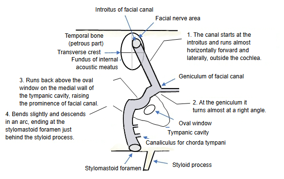
Here the course of the facial canal is divided into four steps.
| 1 | Forward and lateral | The canal begins at the introitus of the facial canal in the facial nerve area (the upper front of the four areas of the fundus of the internal acoustic meatus, above the transverse crest). From there it runs almost horizontally forward and laterally, above the cochlea. |
| 2 | Bend at almost a right angle | It then turns sharply backward and laterally, almost at a right angle. This bend is the geniculum of the facial canal; the geniculate ganglion lies here. |
| 3 | Over the tympanic cavity | It runs backward and slightly laterally on the medial wall of the tympanic cavity, above the oval window, where it raises a ridge on the wall: the prominence of facial canal. |
| 4 | Down in an arc to the exit | Behind the tympanic cavity it curves downward in an arc and opens just behind the root of the styloid process as the stylomastoid foramen. |
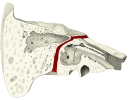 Facial canal (oblique section)
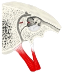 Right tympanic cavity (lateral view, section)
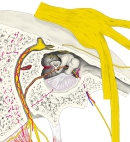 Right tympanic cavity with nerves
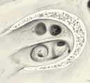 Fundus of the right internal acoustic meatus
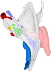 Right temporal bone and ear (superior view)
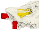 Nerves at the fundus
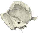 Left temporal bone (medial surface)
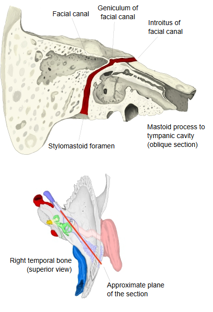
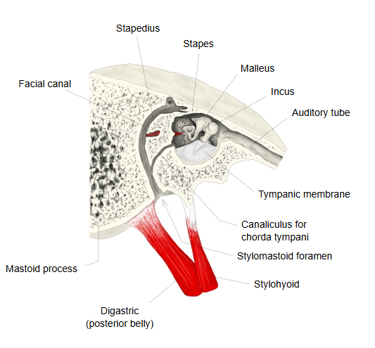
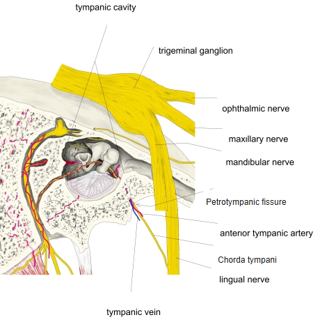
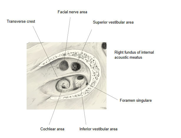
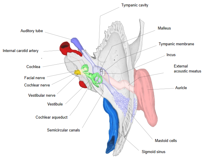
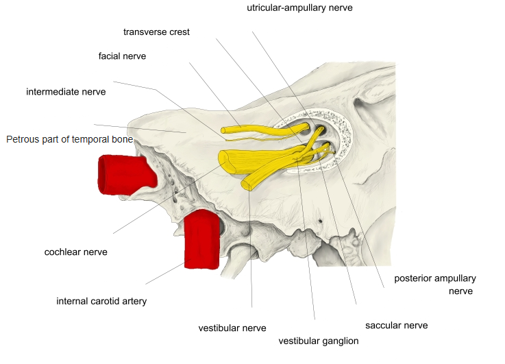
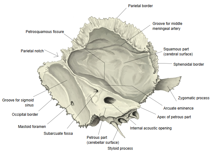
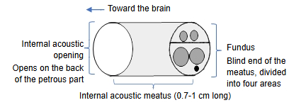
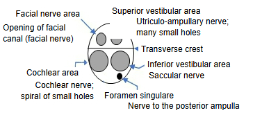
The facial canal is the path of the facial nerve: it begins at the facial nerve area above the transverse crest of the fundus of the internal acoustic meatus and ends at the stylomastoid foramen. At its first bend, the geniculum (see geniculum of facial nerve), the nerve gives off one branch, the greater petrosal nerve, which runs forward through the canal for the greater petrosal nerve, leaves the bone at the hiatus for the greater petrosal nerve on the front of the petrous part and continues in the groove for the greater petrosal nerve. In the descending part the canal gives off the canaliculus for the chorda tympani, which carries the chorda tympani into the tympanic cavity. The bony wall is thin over the tympanic cavity and may be partly missing, so the nerve can be exposed in middle-ear disease and surgery.
Segments
Labyrinthine segment: from the fundus of the internal acoustic meatus to the geniculum, above the cochlea. Its beginning is the narrowest part of the canal.
Tympanic segment: from the geniculum to the second bend, along the medial wall of the tympanic cavity, below the lateral semicircular canal and above the oval window.
Mastoid segment: from the second bend straight down, along the posterior wall of the tympanic cavity, to the stylomastoid foramen.
Reference websites
[Wikipedia]
· Facial canal, Wikipedia.
· Facial nerve, Wikipedia.
[Websites with illustrations]
· The facial nerve (CN VII) (TeachMeAnatomy)
· Facial nerve: origin, function, branches and anatomy (Kenhub)
Related terms
- Facial nerve
- Introitus of facial canal
- Prominence of facial canal
- Geniculum of facial nerve
- Stylomastoid foramen
- Internal acoustic meatus
- Petrous part
References and sources
- Further reading: Facial canal, Wikipedia.
- Further reading: Facial nerve, Wikipedia.
- Federative International Programme for Anatomical Terminology (FIPAT). Terminologia Anatomica, 2nd ed. (TA2), 2019.