Renal hilum (Latin: hilum renale)
Also called the hilum of kidney
Overview
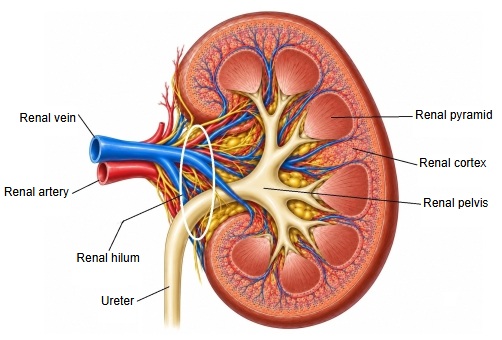
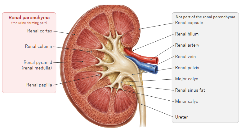
· In short, it is the place where the renal vessels and the ureter enter and leave the kidney.
· From front to back, the structures at the hilum are usually the renal vein, the renal artery and the renal pelvis, which continues as the ureter. Together these structures form the renal pedicle.
· The hilum opens into a recess surrounded by the renal parenchyma, the renal sinus, which holds the renal pelvis, the renal calyces and the vessels; the spaces between them are filled with fat.
· The hilum lies at about the level of the first to second lumbar vertebrae (sources differ). The right kidney sits slightly lower than the left because of the liver.
Reference websites
[Wikipedia]
· Renal hilum, Wikipedia.
· Kidney, Wikipedia.
[Websites with illustrations]
· Gross anatomy of the kidney (Lumen Learning, Anatomy and Physiology II)
· Kidney anatomy (Blausen) (Wikimedia Commons)

· The anatomy of the kidney (Interactive Biology)
Related terms
References and sources
- Further reading: Renal hilum, Wikipedia.
- Further reading: Kidney, Wikipedia.
- Federative International Programme for Anatomical Terminology (FIPAT). Terminologia Anatomica, 2nd ed. (TA2), 2019.