Renal papilla (Latin: papilla renalis)

Overview

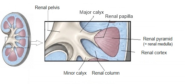
The renal papilla is the part at the tip of a renal pyramid where the papillary ducts gather in numbers and which projects into a minor calyx.
Renal pyramid = renal medullaPapillary duct: the last part of the collecting ductAbout 20 on average
Each renal pyramid usually has one renal papilla, but in some cases several pyramids join to form one renal papilla.
Compound papilla

· Compound papilla: the tips of two or three renal pyramids may fuse into one renal papilla, so there are fewer papillae than pyramids. Compound papillae are common at the upper and lower ends of the kidney.

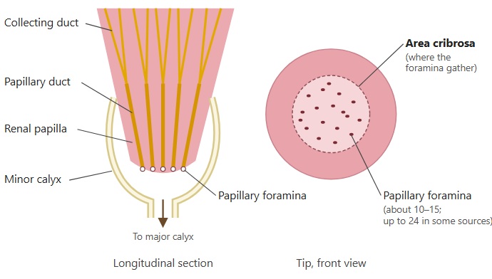
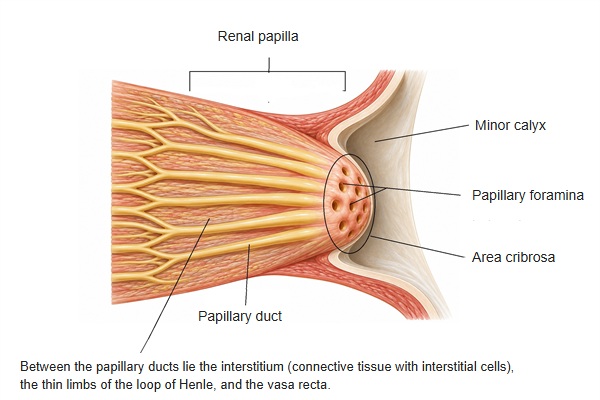
· Seen from the surface, the renal papilla has many openings, the papillary foramina (about 10 to 15; some sources give up to 24), so that it looks like a sieve even to the naked eye; this area is called the area cribrosa.

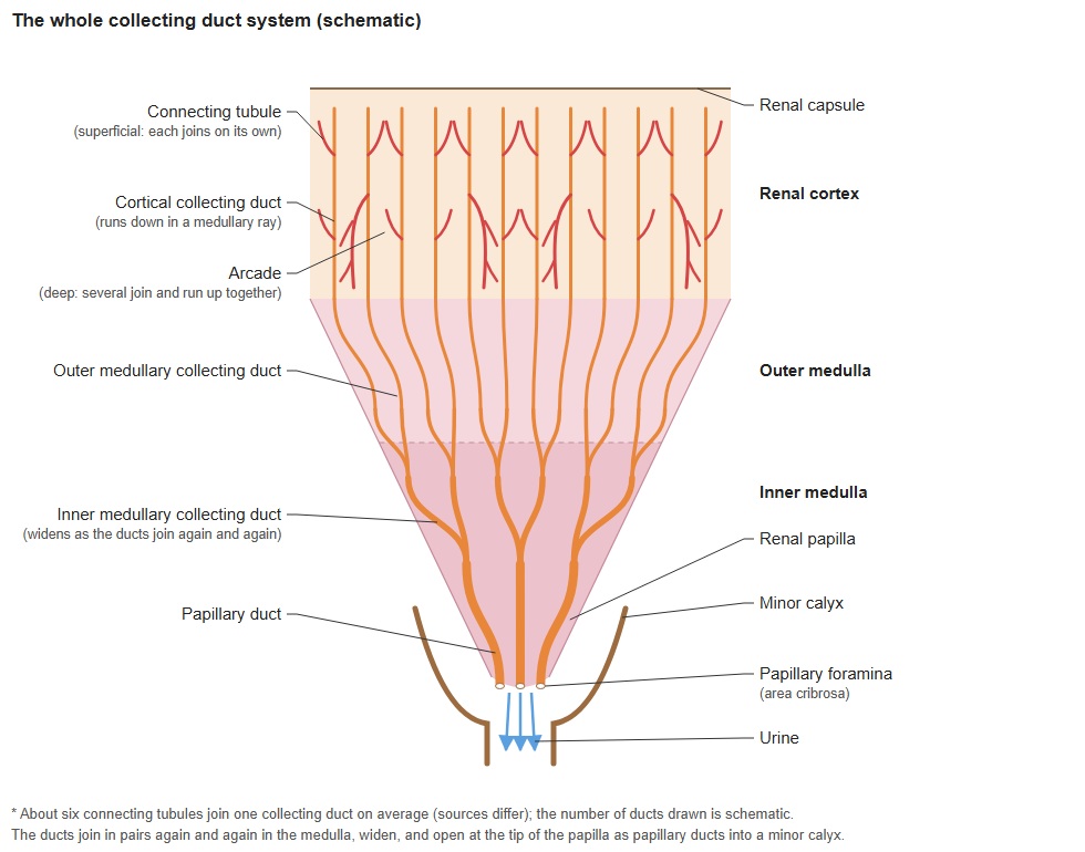
· The medullary collecting ducts join to form the papillary ducts. Through the papillary foramina the primary urine leaves the renal medulla and enters the minor calyx, where it becomes urine.

· The transitional epithelium (urothelium) that lines the urinary tract begins near the renal papilla.

· Drugs such as painkillers (NSAIDs) can cause necrosis of the cells of the papilla (papillary necrosis). Lesions of the papilla are also linked to kidney stones.
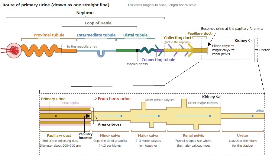
Route of primary urine drawn as one straight line, from the renal corpuscle through the proximal, intermediate and distal tubules, the connecting tubule, collecting duct and papillary duct; it becomes urine via the papillary foramina and passes the minor calyx, major calyx and renal pelvis to the ureter. Below, the end enlarged: papillary duct in the renal papilla, papillary foramina and area cribrosa, then minor calyx, major calyx, renal pelvis and ureter, each with a short note.
(click to enlarge)
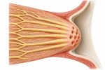
Section of a kidney with part of it enlarged: a renal papilla, the tip of a renal pyramid (renal medulla), projects into a minor calyx; the minor calyces join the major calyx and the renal pelvis; renal cortex and renal column are labelled.
(click to enlarge)
Left, longitudinal section: collecting ducts join to form papillary ducts that open as papillary foramina at the tip of the renal papilla, which projects into a minor calyx leading to a major calyx. Right, the tip seen from the front: the area cribrosa, where the papillary foramina (about 10 to 15; up to 24 in some sources) gather.
(click to enlarge)
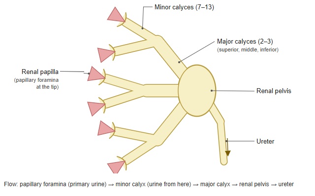
  • Small picture of the renal calyces, renal pelvis and ureter; opens the labelled figure.Renal calyces
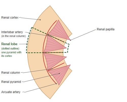
  • Small picture of a renal lobe: one renal pyramid with the cortex around it; opens the labelled figure.Renal lobe
  • Small picture of the whole collecting duct system from the cortex to the minor calyx; opens the labelled figure.The whole collecting duct
  • Small picture of a renal papilla with its papillary ducts opening into a minor calyx; opens the labelled figure.Renal papilla

Reference websites

[Wikipedia]

· Renal papilla, Wikipedia.

· Kidney, Wikipedia.

[Websites with illustrations]

· Gross anatomy of the kidney (Lumen Learning, Anatomy and Physiology II)

· Kidney cross-section (EBM Consult)

· Kidney anatomy (Blausen) (Wikimedia Commons)

· Hydronephrosis (ultrasound) (NYSORA)

References and sources

  1. Further reading: Renal papilla, Wikipedia.
  2. Further reading: Kidney, Wikipedia.
  3. Federative International Programme for Anatomical Terminology (FIPAT). Terminologia Anatomica, 2nd ed. (TA2), 2019.