Posterior cricoarytenoid (Latin: musculus cricoarytenoideus posterior)
- Overview
- Illustrations
- Origin / insertion
- Action
- Nerve / blood supply
- Reference websites
- (Wikipedia, websites with illustrations)
- Related terms
Overview
- Superficial muscles of the neck
- Suprahyoid and infrahyoid muscles
- Deep muscles of the neck
- Muscles of the pharynx
- Intrinsic laryngeal muscles (9) : cricothyroid, posterior cricoarytenoid, lateral cricoarytenoid, thyroarytenoid, vocalis, transverse arytenoid, oblique arytenoid, aryepiglotticus, thyroepiglotticus
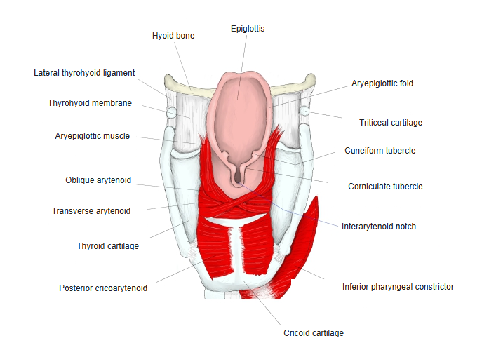
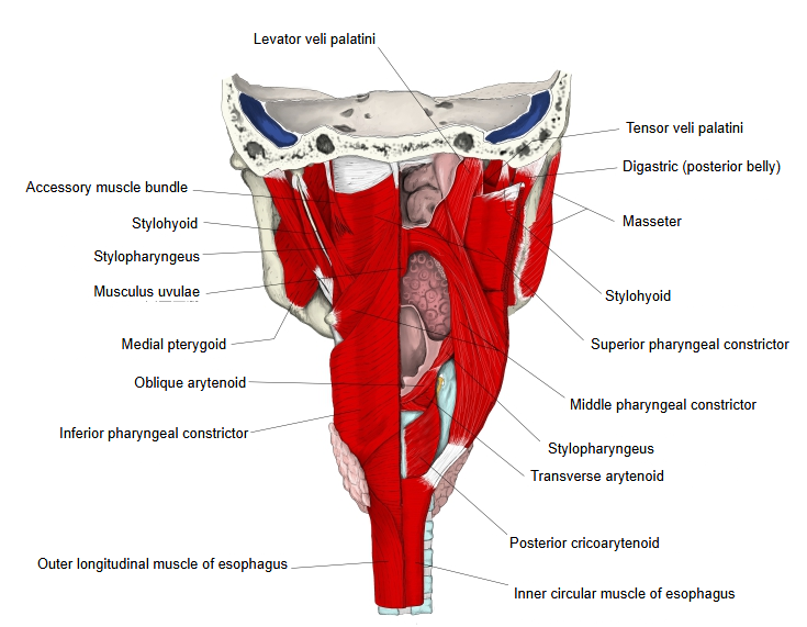
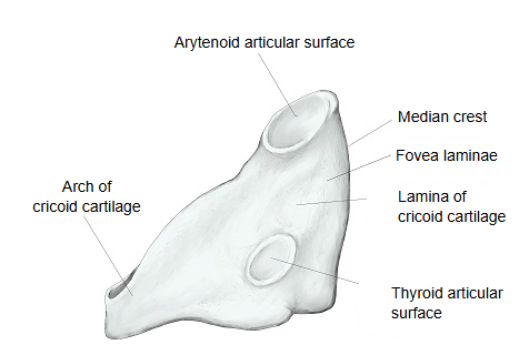
· A paired, fan-shaped muscle on the back of the lamina of the cricoid cartilage, just in front of the laryngeal part of the pharynx.
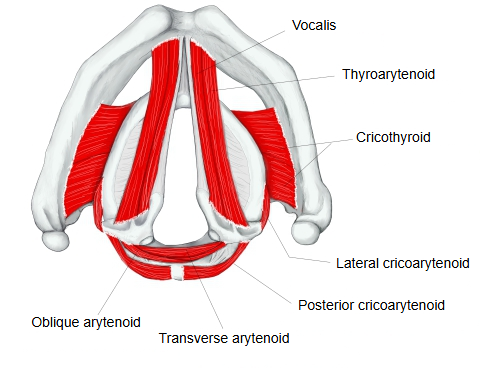
· It is the only abductor of the vocal folds. If both muscles are paralysed (for example after injury to both recurrent laryngeal nerves), the vocal folds stay near the midline and breathing can be blocked.
· Small extra slips are sometimes present: from the lower border of the cricoid lamina to the inferior horn of the thyroid cartilage (the ceratocricoid muscle, musculus ceratocricoideus), and from the inferior horn to the muscular process of the arytenoid cartilage (sometimes called the ceratoarytenoid). How often they occur: sources differ.
The intrinsic laryngeal muscles at a glance (attachments simplified):
| Muscle | Origin | Insertion | Main action |
|---|---|---|---|
| Cricothyroid | front and side of the arch of the cricoid cartilage | lower border and inferior horn of the thyroid cartilage | tilts the thyroid cartilage forward: lengthens and tenses the vocal folds (the only one supplied by the superior laryngeal nerve) |
| Posterior cricoarytenoid | back of the lamina of the cricoid cartilage | back of the muscular process of the arytenoid cartilage | abducts the vocal folds: the only muscle that opens the rima glottidis |
| Lateral cricoarytenoid | upper border of the arch of the cricoid cartilage | front of the muscular process of the arytenoid cartilage | adducts the vocal folds |
| Thyroarytenoid | inner surface of the thyroid cartilage near the angle; cricothyroid ligament | anterolateral surface and muscular process of the arytenoid cartilage | shortens and relaxes the vocal folds; helps to close the glottis |
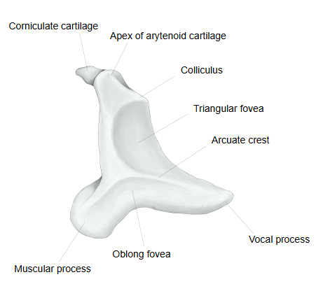
| Vocalis (deep part) | inner surface of the thyroid cartilage near the angle | vocal process and oblong fovea of the arytenoid cartilage | fine-tunes the tension of the vocal fold |
| Thyroepiglotticus (part) | with the thyroarytenoid | aryepiglottic fold and margin of the epiglottis | widens the laryngeal inlet |
| Transverse arytenoid | back and lateral border of one arytenoid cartilage | the same parts of the other arytenoid cartilage | draws the arytenoid cartilages together |
| Oblique arytenoid | back of the muscular process of one arytenoid cartilage | apex of the other arytenoid cartilage | draws the arytenoid cartilages together; narrows the laryngeal inlet |
| Aryepiglotticus (part) | continuation of the oblique arytenoid at the apex of the arytenoid cartilage | side of the epiglottis, through the aryepiglottic fold | narrows the laryngeal inlet |
Illustrations
More figures: Multi-muscle illustrations: Neck · Pharynx and esophagus (posterior view) · Muscles of the neck (list) · Larynx
Origin / insertion
[Origin] : the shallow depression (fovea laminae) on the back of the lamina of the cricoid cartilage, beside the median ridge.
The fibres run upward and laterally and converge on the arytenoid cartilage; the upper fibres are almost horizontal, the lower ones almost vertical.
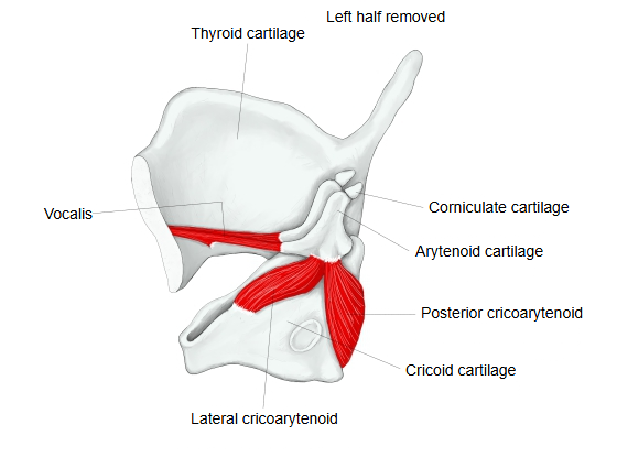
[Insertion] : the back of the muscular process of the arytenoid cartilage.
Action
Pulls the muscular process backward and medially. The arytenoid cartilage turns outward on the cricoarytenoid joint, its vocal process swings laterally, and the vocal folds move apart: the rima glottidis opens. It contracts with every breath in, and it also steadies the arytenoid cartilage while the cricothyroid tenses the vocal folds.
Nerve / blood supply
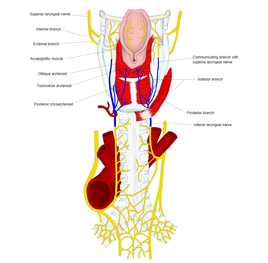
· Nerve : recurrent laryngeal nerve (its end part is called the inferior laryngeal nerve), a branch of the vagus nerve (cranial nerve X).
· Arteries : laryngeal branches of the superior laryngeal artery (from the superior thyroid artery) and the inferior laryngeal artery (from the inferior thyroid artery).
· Veins : the superior laryngeal vein to the superior thyroid vein, and the inferior laryngeal veins to the inferior thyroid veins.
 Laryngeal nerves (posterior view)
Reference websites
[Wikipedia]
· Posterior cricoarytenoid muscle, Wikipedia.
[Websites with illustrations]
· Posterior cricoarytenoid (Kenhub)
Related terms
- Intrinsic laryngeal muscles
- Cricoid cartilage
- Arytenoid cartilage
- Rima glottidis
- Lateral cricoarytenoid
- Cricothyroid
- Recurrent laryngeal nerve
- Larynx
References and sources
- FIPAT. Terminologia Anatomica, 2nd ed. (TA2), 2019: musculus cricoarytenoideus posterior.
- Posterior cricoarytenoid muscle, Wikipedia.