Thyroarytenoid (Latin: musculus thyroarytenoideus)
- Overview
- Illustrations
- Origin / insertion
- Action
- Nerve / blood supply
- Reference websites
- (Wikipedia, websites with illustrations)
- Related terms
Overview
- Superficial muscles of the neck
- Suprahyoid and infrahyoid muscles
- Deep muscles of the neck
- Muscles of the pharynx
- Intrinsic laryngeal muscles (9) : cricothyroid, posterior cricoarytenoid, lateral cricoarytenoid, thyroarytenoid, vocalis, transverse arytenoid, oblique arytenoid, aryepiglotticus, thyroepiglotticus
· A broad, thin sheet in the side wall of the larynx, lateral to the vocal fold, the laryngeal ventricle and the laryngeal saccule.
· Its deep, medial part beside the vocal ligament is the vocalis. Sources differ: TA2 lists the vocalis as a muscle of its own, while many texts describe it as part of the thyroarytenoid.
· Upper fibres that continue into the aryepiglottic fold form the thyroepiglotticus; a few fibres along the wall of the ventricle are sometimes called the ventricularis.
The intrinsic laryngeal muscles at a glance (attachments simplified):
| Muscle | Origin | Insertion | Main action |
|---|---|---|---|
| Cricothyroid | front and side of the arch of the cricoid cartilage | lower border and inferior horn of the thyroid cartilage | tilts the thyroid cartilage forward: lengthens and tenses the vocal folds (the only one supplied by the superior laryngeal nerve) |
| Posterior cricoarytenoid | back of the lamina of the cricoid cartilage | back of the muscular process of the arytenoid cartilage | abducts the vocal folds: the only muscle that opens the rima glottidis |
| Lateral cricoarytenoid | upper border of the arch of the cricoid cartilage | front of the muscular process of the arytenoid cartilage | adducts the vocal folds |
| Thyroarytenoid | inner surface of the thyroid cartilage near the angle; cricothyroid ligament | anterolateral surface and muscular process of the arytenoid cartilage | shortens and relaxes the vocal folds; helps to close the glottis |
| Vocalis (deep part) | inner surface of the thyroid cartilage near the angle | vocal process and oblong fovea of the arytenoid cartilage | fine-tunes the tension of the vocal fold |
| Thyroepiglotticus (part) | with the thyroarytenoid | aryepiglottic fold and margin of the epiglottis | widens the laryngeal inlet |
| Transverse arytenoid | back and lateral border of one arytenoid cartilage | the same parts of the other arytenoid cartilage | draws the arytenoid cartilages together |
| Oblique arytenoid | back of the muscular process of one arytenoid cartilage | apex of the other arytenoid cartilage | draws the arytenoid cartilages together; narrows the laryngeal inlet |
| Aryepiglotticus (part) | continuation of the oblique arytenoid at the apex of the arytenoid cartilage | side of the epiglottis, through the aryepiglottic fold | narrows the laryngeal inlet |
Illustrations
More figures: Multi-muscle illustrations: Neck · Muscles of the neck (list) · Larynx
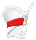 Thyroarytenoid
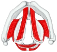 Intrinsic laryngeal muscles (superior view)
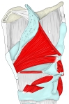 Intrinsic laryngeal muscles (lateral view)
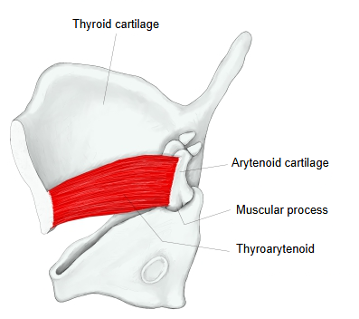
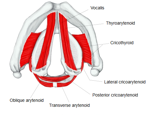
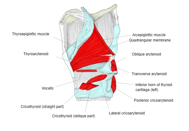
Origin / insertion
[Origin] : the inner surface of the thyroid cartilage near the midline (the lower half of the angle) and the adjacent cricothyroid ligament.
From the origin the fibres run almost horizontally backward and laterally.
[Insertion] : the anterolateral surface of the arytenoid cartilage, from the vocal process to the muscular process, including the oblong fovea.
Sources differ on how far up the insertion reaches: some texts give only the lower part (muscular process and oblong fovea), others most of the anterolateral surface.
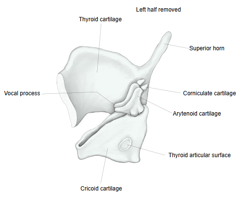
Action
Draws the arytenoid cartilages forward toward the thyroid cartilage, so the vocal folds become shorter and slacker (the opposite of the cricothyroid). Its lateral fibres turn the arytenoid cartilage inward and help to close the rima glottidis; its upper fibres help to narrow the laryngeal vestibule.
Nerve / blood supply
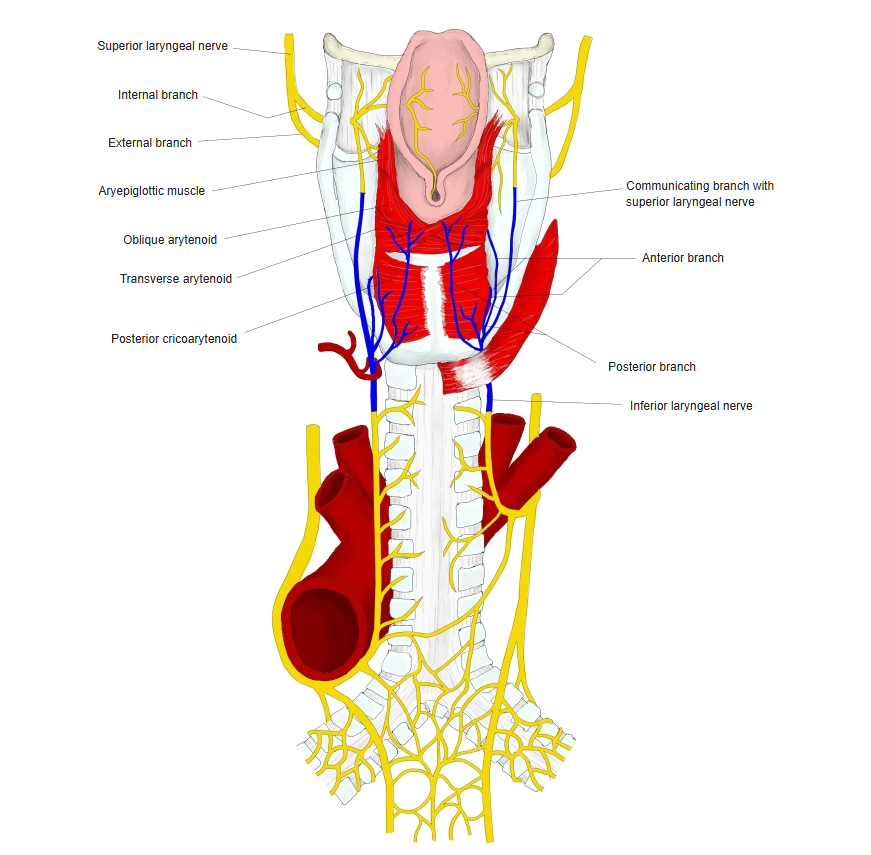
· Nerve : recurrent laryngeal nerve (its end part is called the inferior laryngeal nerve), a branch of the vagus nerve (cranial nerve X).
· Arteries : laryngeal branches of the superior laryngeal artery (from the superior thyroid artery) and the inferior laryngeal artery (from the inferior thyroid artery).
· Veins : the superior laryngeal vein to the superior thyroid vein, and the inferior laryngeal veins to the inferior thyroid veins.
 Laryngeal nerves (posterior view)
Reference websites
[Wikipedia]
· Thyroarytenoid muscle, Wikipedia.
[Websites with illustrations]
· Thyroarytenoid (Kenhub)
Related terms
- Intrinsic laryngeal muscles
- Thyroid cartilage
- Arytenoid cartilage
- Vocal fold
- Vocalis
- Thyroepiglotticus
- Cricothyroid
- Vocal ligament
- Recurrent laryngeal nerve
References and sources
- FIPAT. Terminologia Anatomica, 2nd ed. (TA2), 2019: musculus thyroarytenoideus.
- Thyroarytenoid muscle, Wikipedia.