Urinary tract
Overview
Primary urine becomes urine where it leaves the papillary foramina into the minor calyx.
· The urinary tract begins with the renal calyces (minor and major calyces) and renal pelvis inside the kidney. The ureter carries the urine to the urinary bladder, which stores it until it leaves the body through the urethra. This page covers the passages only; for the system as a whole see the urinary system.
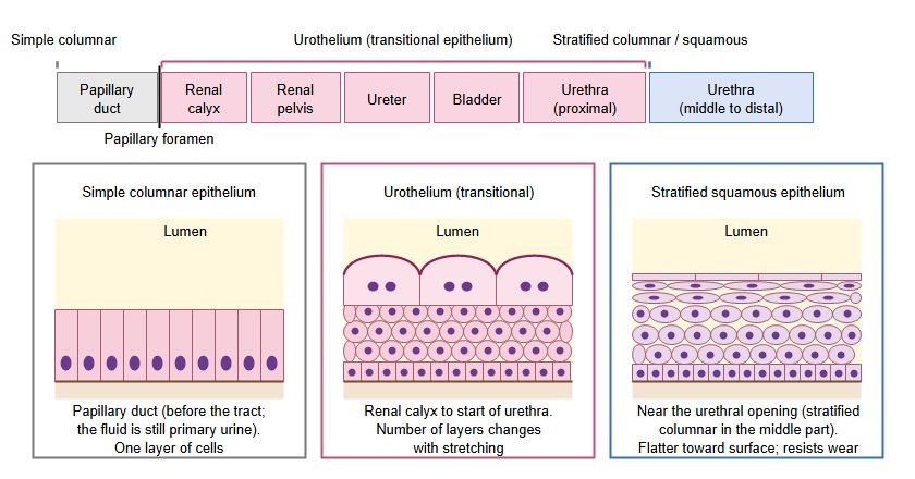
· The inner surface from the calyces to the start of the urethra is lined by urothelium (transitional epithelium), which changes shape as the wall stretches and relaxes.
· The fluid in the collecting ducts and papillary ducts of the kidney is primary urine. It becomes urine where it leaves the papillary foramina of the renal papilla into the minor calyx. Its composition hardly changes on the way through the urinary tract.
· Smooth muscle in the walls of the calyces, renal pelvis and ureter contracts in waves (peristalsis) and moves the urine to the bladder. The ureter passes obliquely through the bladder wall, so as the filling bladder stretches, the ureter is pressed shut and urine does not easily flow back.
Wall of the urinary tract
· From the inside, the wall consists of mucosa, muscle layer and adventitia (the upper surface of the bladder is covered by peritoneum).
· The surface of the mucosa is urothelium (transitional epithelium). When urine collects and the wall stretches, its cells flatten and the layer becomes thinner; when the wall contracts it becomes thicker. It also keeps the constituents of urine from soaking into the wall.
· The muscle layer is smooth muscle. In the bladder it is called the detrusor muscle, which contracts to push the urine out. The urethra has an internal urethral sphincter and an external urethral sphincter.
Reference websites
[Wikipedia]
· Urinary system, Wikipedia.
· Transitional epithelium, Wikipedia.
Related terms
References and sources
- Further reading: Urinary system, Wikipedia.
- Federative International Programme for Anatomical Terminology (FIPAT). Terminologia Anatomica, 2nd ed. (TA2), 2019.