Proximal epiphysis of tibia (Latin: epiphysis proximalis tibiae)
Also called the upper end of tibia
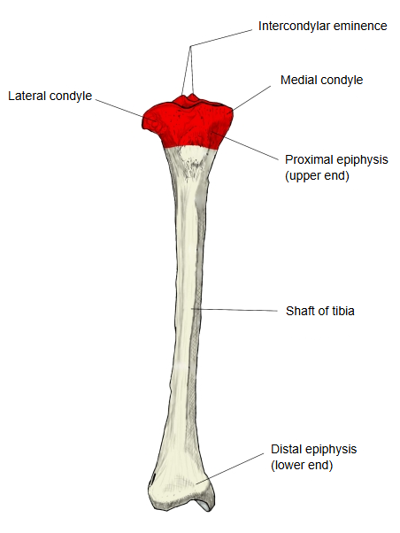
When the tibia is divided into three parts, the proximal epiphysis is the expanded upper part; it is also called the upper end of the tibia.
- Proximal epiphysis : much wider than the shaft. = upper end
- Shaft of tibia : the long middle part of the bone.
- Distal epiphysis : wider than the shaft, but less than the upper end. = lower end
The upper end has a medial condyle and a lateral condyle, which spread out to either side. The upper surfaces of both condyles are covered with cartilage; these are the superior articular surfaces, which articulate with the medial and lateral condyles of the femur. The articular capsule is attached around them. From the middle of the articular surface a ridge rises upwards, the intercondylar eminence; its two ends form small tubercles, the medial and lateral intercondylar tubercles. In front of the eminence lies the anterior intercondylar area and behind it the posterior intercondylar area; both give attachment to ligaments. Behind the lateral condyle, below the margin of the joint surface, is a shallow oval joint surface, the fibular articular facet, which articulates with the head of the fibula. On the back and medial side of the medial condyle is a groove for the tendon of semimembranosus.
· Each superior articular surface carries a meniscus; the anterior and posterior intercondylar areas give attachment to the cruciate ligaments and to the horns of the menisci.
· At the front, just below the condyles, the tibial tuberosity receives the patellar ligament.
The following features are found on it.
| 1 | Superior articular surfaces | facies articularis superior | 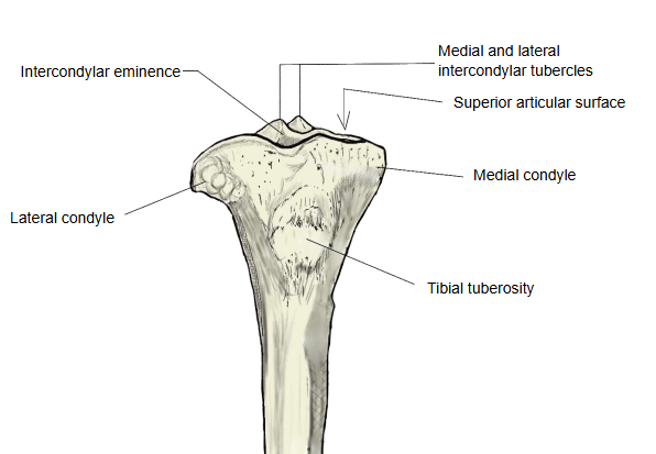 |
| 2 | Intercondylar eminence | eminentia intercondylaris | |
| 3 | Medial and lateral intercondylar tubercles | tuberculum intercondylare mediale / laterale | |
| 4 | Anterior and posterior intercondylar areas | area intercondylaris anterior / posterior | |
| 5 | Medial and lateral condyles | condylus medialis / lateralis |
Attachments
| Muscles | 1 | Fibularis longus (origin: inconstant, a few fibres from the lateral condyle; it arises mainly from the fibula) | 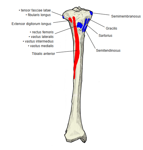 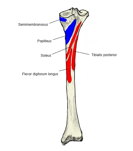 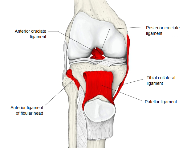 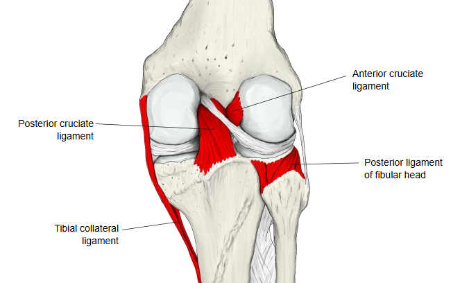 |
| 2 | Tensor fasciae latae (insertion, through the iliotibial tract) | ||
| 3 | Semimembranosus (insertion) | ||
| Ligaments | 1 | Tibial collateral ligament | |
| 2 | Anterior cruciate ligament | ||
| 3 | Posterior cruciate ligament | ||
| 4 | Anterior ligament of fibular head (see ligaments of fibular head) | ||
| 5 | Posterior ligament of fibular head |
Reference websites
[Wikipedia]
· Tibia, Wikipedia.
· Knee, Wikipedia.
[Websites with illustrations]
· Upper surface of the right tibia (Gray's Anatomy, fig. 257) (Wikimedia Commons)

· Bones of the right leg, anterior surface (Gray's Anatomy, fig. 258) (Wikimedia Commons)

· The tibia (TeachMeAnatomy)
· Tibia (Kenhub)
Where it is found
Related terms
- Shaft of tibia
- Distal epiphysis of tibia
- Medial condyle of tibia
- Lateral condyle of tibia
- Intercondylar eminence
- Tibial tuberosity
- Fibular articular facet