Cranial synchondroses (Latin: synchondroses cranii)
Also called the synchondroses of skull or the cranial synchondrosis
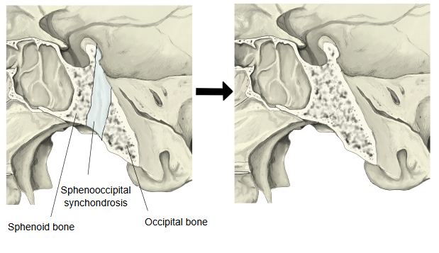
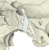 Sphenooccipital synchondrosis
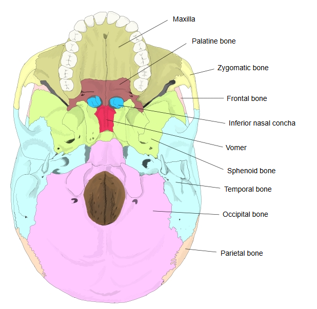
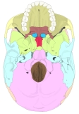 External cranial base
In the usual classification of joints shown below, the cranial synchondroses are "hyaline cartilage joints" (synchondroses) among the cartilaginous joints of the fixed (immovable) joints, unlike the synovial joints, which have a joint cavity.
of joints
- Fixed (immovable) joints
- a. Fibrous joints : syndesmosis, suture, gomphosis.
- b. Cartilaginous joints : fibrocartilage joint (symphysis), hyaline cartilage joint (synchondrosis).
- c. Synostosis : bones fused by bone (a joint that has turned to bone).
- Movable joints : there is a space (the joint cavity) between the bones, which are completely separate: the "joints in the narrow sense" (synovial joints).
The hyaline cartilage joint is thus one of the cartilaginous joints among the fixed joints.
When a text speaks of a cartilaginous joint without saying which kind, it usually means a hyaline cartilage joint (synchondrosis).
Most of the cartilaginous joints of the skull disappear as ossification goes on. They are the remains of the cartilage of the embryonic cartilaginous skull (the chondrocranium), left between the centres of ossification of the bones of the cranial base. While they last, they act as growth plates for the cranial base; most ossify during childhood or adolescence, so in the adult the bones are fused. Some never fully ossify: the sphenopetrosal and petro-occipital synchondroses persist as cartilage-filled fissures.
One classical textbook lists the following six cranial synchondroses.
| Name | Notes | |
| 1 | Sphenooccipital synchondrosis | The cartilaginous joint between the basilar part of the occipital bone (basilar part) and the body of the sphenoid; it ossifies after puberty (by about the age of 20, according to this textbook). |
| 2 | Sphenopetrosal synchondrosis | Fills the sphenopetrosal fissure, between the greater wing of the sphenoid bone and the petrous part of the temporal bone. |
| 3 | Petro-occipital synchondrosis | Lies in the petro-occipital fissure; the inferior petrosal sinus runs along it. |
| 4 | Posterior intra-occipital synchondrosis (between the squama and the lateral parts of the occipital bone) | Seen in infants up to about the second year of life: they separate each lateral part of the occipital bone from the basilar part in front and from the occipital squama behind, around the foramen magnum. |
| 5 | Anterior intra-occipital synchondrosis (between the basilar part and the lateral parts of the occipital bone) | |
| 6 | Intersphenoidal synchondrosis | Present only in the fetus, up to the end of the seventh month; it runs across the body of the sphenoid near the tuberculum sellae, between its front and back parts. |
Another source lists the following four cranial synchondroses, with the ages at which they ossify:
1. 1. Sphenoethmoidal synchondrosis: between 5 and 20 years
2. 2. Intersphenoidal synchondrosis: from birth to 7 years (the textbook above says it exists only before birth; sources differ)
3. 3. Sphenooccipital synchondrosis: at 14–15 years in males and 12–13 years in females
4. 4. Intra-occipital synchondroses: by 3–5 years. They ossify by endochondral ossification and take part in the growth of the cranial base.
English anatomical dictionaries define the cranial synchondroses as the cartilaginous joints of the skull and list:
· sphenoethmoidal synchondrosis,
· sphenooccipital synchondrosis,
· sphenopetrosal synchondrosis,
· petro-occipital synchondrosis,
· anterior intra-occipital synchondrosis,
· posterior intra-occipital synchondrosis.
Cranial synchondroses form where separate centres of ossification appear within one continuous mass of hyaline cartilage. As the neighbouring centres grow towards each other, the cartilage left between them works as a growing spacer while the bones develop; in most places it is finally replaced by bone and the two bones fuse. In the skull these joints lie in the developing chondrocranium, which gives the first support to the rapidly growing brain; they are found within and between the occipital, temporal, sphenoid and ethmoid bones. Sources differ on which joints are listed and on the age at which each one closes.
Related terms
- Cranial fossa
- Cranial cavity
- Cranial base
- Basilar part of occipital bone
- Anterior cranial fossa
- Middle cranial fossa
- Sphenooccipital synchondrosis
- Cartilaginous joint
- Clivus
- Foramen lacerum
- External cranial base
- Sphenoid bone
- Occipital bone
References and sources
- Further reading: Synchondrosis, Wikipedia.
- Further reading: Spheno-occipital synchondrosis, Wikipedia.
- Federative International Programme for Anatomical Terminology (FIPAT). Terminologia Anatomica, 2nd ed. (TA2), 2019.