Ethmoidal bulla (Latin: bulla ethmoidalis)
Also called the ethmoid bulla
· Classic anatomy texts (and Wikipedia) describe it as formed by the middle ethmoidal cells, whereas surgical texts usually treat it as the largest anterior ethmoidal cell. It is a key landmark in endoscopic sinus surgery.
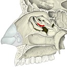 Right nasal cavity, lateral wall (conchae removed)
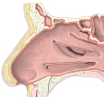 Right nasal cavity, lateral wall (conchae cut)
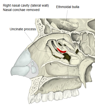
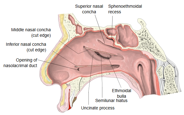
The bulla lies on the lateral wall of the middle nasal meatus, under cover of the middle nasal concha, which has to be lifted or removed to see it.
Structure
The cells inside the bulla open on it or close to it, often just above or below it; the bulla is often the largest of the cells of its group. Just below and in front of it is the curved slit of the semilunar hiatus, bounded in front by the uncinate process. The hiatus leads into the ethmoidal infundibulum, which receives the opening of the maxillary sinus and often of the frontal sinus.
Development: the ethmoidal bulla begins to form between the 8th and 12th weeks of gestation.
Variation: its size follows that of the cells it contains; it may be a large air cell or little more than a bony ridge in the middle meatus.
Reference websites
[Wikipedia]
· Ethmoid bulla, Wikipedia.
[Websites with illustrations]
· Lateral wall of the nasal cavity, conchae removed (Gray's Anatomy) (Wikimedia Commons)

· The paranasal sinuses (TeachMeAnatomy)
· Nasal cavity (Kenhub)
Where it is found
Related terms
- Ethmoidal air cells
- Uncinate process
- Middle nasal concha
- Middle nasal meatus
- Ethmoidal labyrinth
- Maxillary hiatus
References and sources
- Further reading: Ethmoid bulla, Wikipedia.
- Further reading: Ethmoid sinus, Wikipedia.
- Federative International Programme for Anatomical Terminology (FIPAT). Terminologia Anatomica, 2nd ed. (TA2), 2019.