Laryngeal lymphoid nodules
Also called Laryngeal lymph follicles
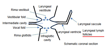
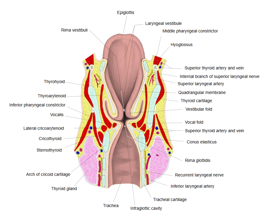
In the lamina propria of the mucosa of the larynx, lymphoid nodules are found here and there; those in the laryngeal ventricle are called the laryngeal lymphoid nodules.
Laryngeal mucosa = surface epithelium + lamina propriaLarynx: part of the airway of the respiratory systemLymphoid nodules: spherical clusters of lymphocytesLaryngeal ventricle: the space that bulges outwards on each side from the laryngeal cavity
A Japanese anatomy textbook describes them as follows.
The lamina propria of the laryngeal mucosa is rich in elastic fibres, and free cells such as lymphocytes, mast cells, eosinophils and plasma cells are scattered in it. Here and there it contains lymphoid nodules; those in the laryngeal ventricle in particular are called the laryngeal lymphoid nodules.
· They are part of the lymphoid tissue that lines the airway and takes part in local immune defence, like the lymphoid tissue of the tonsils (compare the lymph nodes, which are organs with a capsule). Sources differ in naming: some call the collection in the ventricle the "laryngeal tonsil".
Related terms
References and sources
- Further reading: Larynx, Wikipedia.
- Federative International Programme for Anatomical Terminology (FIPAT). Terminologia Anatomica, 2nd ed. (TA2), 2019.