Lesser wing (Latin: ala minor ossis sphenoidalis)
Also called the lesser wing of the sphenoid
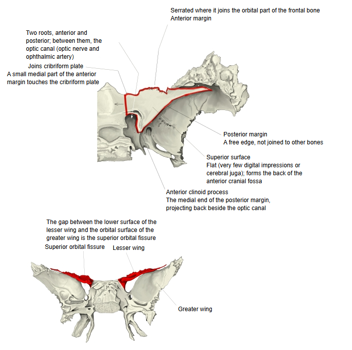
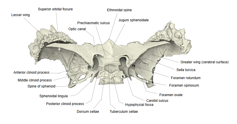
It arises from the body by two roots, anterior and posterior; between them lies the optic canal, which carries the optic nerve and the ophthalmic artery.
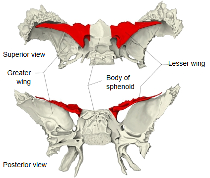
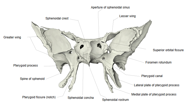
- Body : the central part of the sphenoid; it contains the sphenoidal sinus. Its surfaces are the superior, anterior, inferior and posterior surfaces and two lateral surfaces.
- Processes
- Greater wing : the largest process; it forms part of the middle cranial fossa and of the side wall of the skull.
- Lesser wing : projects to the left and right from the front of the body; it forms part of the anterior cranial fossa.Related terms: optic canal, anterior clinoid process
- Pterygoid process : projects downwards; it consists of two plates, the medial plate and the lateral plate.
Surfaces: the superior surface forms the back part of the anterior cranial fossa and has few digital impressions or cerebral juga. The inferior surface faces the orbit; between it and the greater wing lies the superior orbital fissure.
On the medial side of the superior surface it is continuous with the sphenoidal crest of the body of the sphenoid (the smooth surface between the two lesser wings is the jugum sphenoidale).
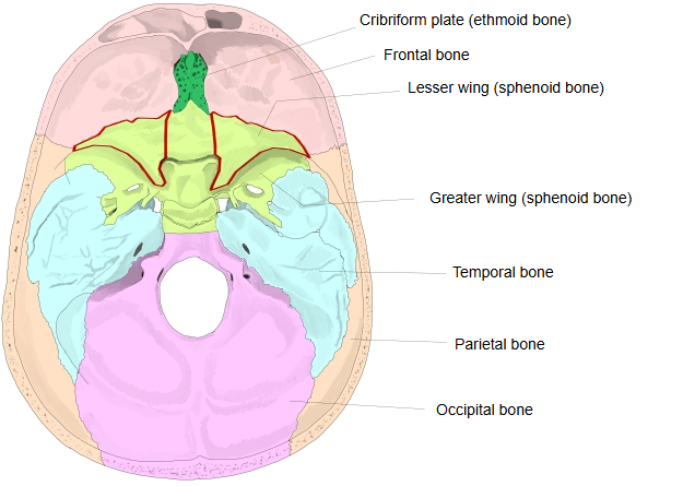
Joins: the whole anterior margin joins the frontal bone (orbital part); a small medial part of the anterior margin joins the ethmoid bone (cribriform plate).
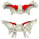 Lesser wing (superior and posterior views)
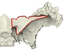 Lesser wing (margins and surfaces)
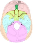 Cranial base (internal surface)
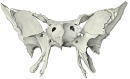 Sphenoid bone (anterior view)
 Sphenoid bone (superior view)
Reference websites
[Wikipedia]
· Lesser wing of sphenoid bone, Wikipedia.
· Sphenoid bone, Wikipedia.
[Websites with illustrations]
· Sphenoid bone: anatomy, function and development (Kenhub)
· Lesser wing of sphenoid bone (superior view) (Wikimedia Commons)

· Sphenoid bone, upper surface (Gray's Anatomy, fig. 145) (Wikimedia Commons)

· Sphenoid bone, upper and posterior surfaces (Gray's Anatomy, fig. 147) (Wikimedia Commons)

Where it is found
Related terms
References and sources
- Further reading: Lesser wing of sphenoid bone, Wikipedia.
- Further reading: Sphenoid bone, Wikipedia.
- Federative International Programme for Anatomical Terminology (FIPAT). Terminologia Anatomica, 2nd ed. (TA2), 2019.