Peritoneal cavity (Latin: cavitas peritonealis)
- Overview
- Reference sites
- Reference websites
- (Wikipedia)
- Compartments
- Where it is found
- Related terms
Overview
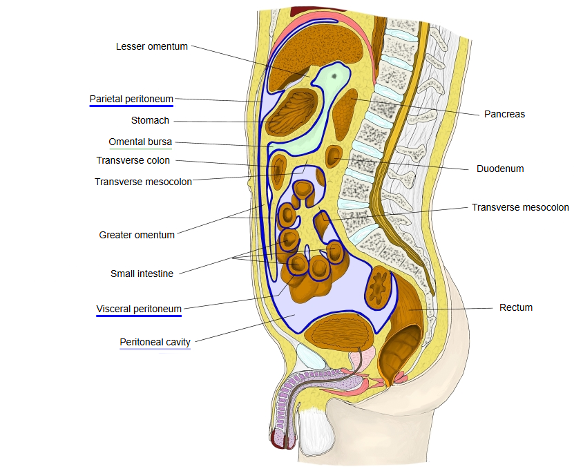
· There is a slight gap between the visceral peritoneum and the parietal peritoneum; this gap is called the peritoneal cavity. Normally it holds only a thin film of serous fluid, which lets the organs slide on each other and on the walls.
· In males it is a closed sac. In females it opens to the outside through the uterine tubes, uterus and vagina.
· An abnormal build-up of fluid in it is called ascites; blood, pus or air in it are signs of serious disease. Sources differ on the normal volume of peritoneal fluid; it is usually given as less than about 50 to 100 mL. Its lowest parts are the rectouterine pouch in females and the rectovesical pouch in males, where fluid tends to collect.
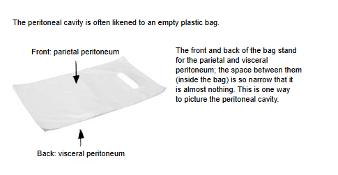
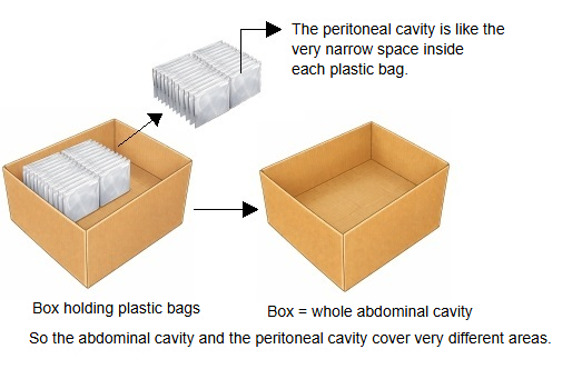
To put it another way, imagine a cardboard box with plastic bags in it. The abdominal cavity is the whole box, while the peritoneal cavity is the narrow space inside each plastic bag.
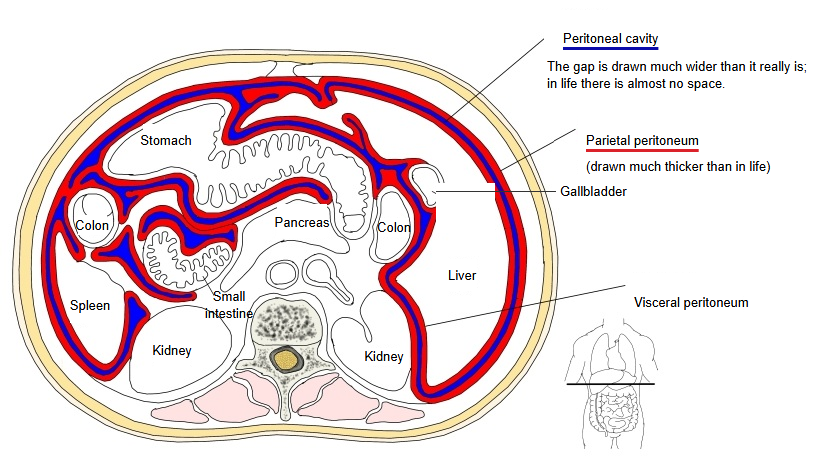
 Abdomen (transverse section, schematic)
 Abdomen and pelvis (median section)
 Body cavities (lateral view)
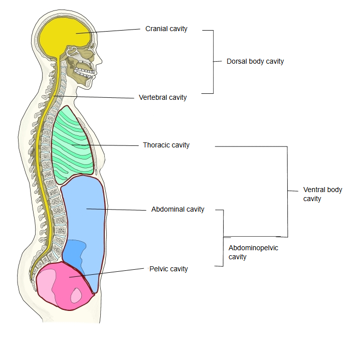
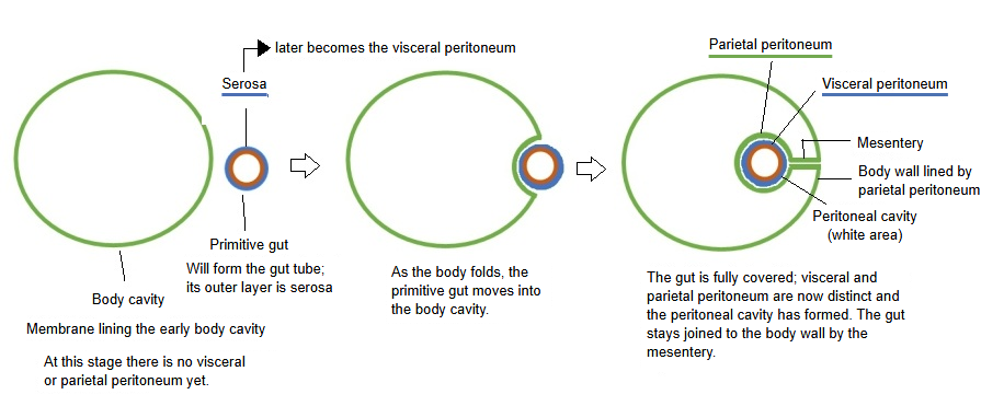
· The peritoneal cavity develops from the part of the embryonic body cavity (intraembryonic coelom) below the diaphragm; the pericardial cavity and the pleural cavities come from the same cavity.
· At first a single serous membrane lines the cavity. As the embryo folds, the gut tube is pushed into the cavity and becomes wrapped in this membrane.
· The membrane on the gut becomes the visceral peritoneum, the membrane on the wall becomes the parietal peritoneum, and the double layer linking them becomes the mesentery.
Reference sites
Reference websites
[Wikipedia]
· Peritoneal cavity, Wikipedia.
· Peritoneum, Wikipedia.
Summary: the peritoneal cavity is the potential space between the parietal peritoneum and the visceral peritoneum; it contains serous fluid, which lets the organs move smoothly against each other.
Compartments
The peritoneal cavity is divided into the greater sac and the lesser sac. The greater sac is the main part of the cavity; the lesser sac (omental bursa) is a smaller pocket behind the stomach and the lesser omentum. The two are connected by the omental foramen (epiploic foramen).
The transverse mesocolon, the mesentery of the transverse colon, divides the greater sac into an upper supracolic compartment and a lower infracolic compartment.
The liver, spleen, stomach and lesser omentum lie in the supracolic compartment. Most of the small intestine, framed by the ascending, transverse and descending colon, lies in the infracolic compartment; the root of the mesentery divides it into right and left spaces, and the paracolic gutters run beside the ascending and descending colon.
Where it is found
Related terms
- Parietal peritoneum
- Visceral peritoneum
- Peritoneum
- Greater omentum
- Transverse mesocolon
- Pleural cavity
- Pericardial cavity
References and sources
- Further reading: Peritoneal cavity, Wikipedia.
- Further reading: Peritoneum, Wikipedia.
- Federative International Programme for Anatomical Terminology (FIPAT). Terminologia Anatomica, 2nd ed. (TA2), 2019.