Pterygomaxillary fissure (Latin: fissura pterygomaxillaris)
Also called the pterygopalatine fissure (older texts)
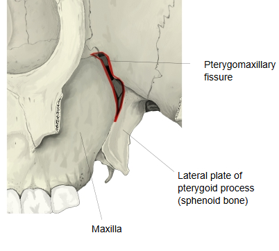
The pterygomaxillary fissure is a gap with some width between the maxilla and the lateral plate of the pterygoid process of the sphenoid bone; it transmits nerves and blood vessels.
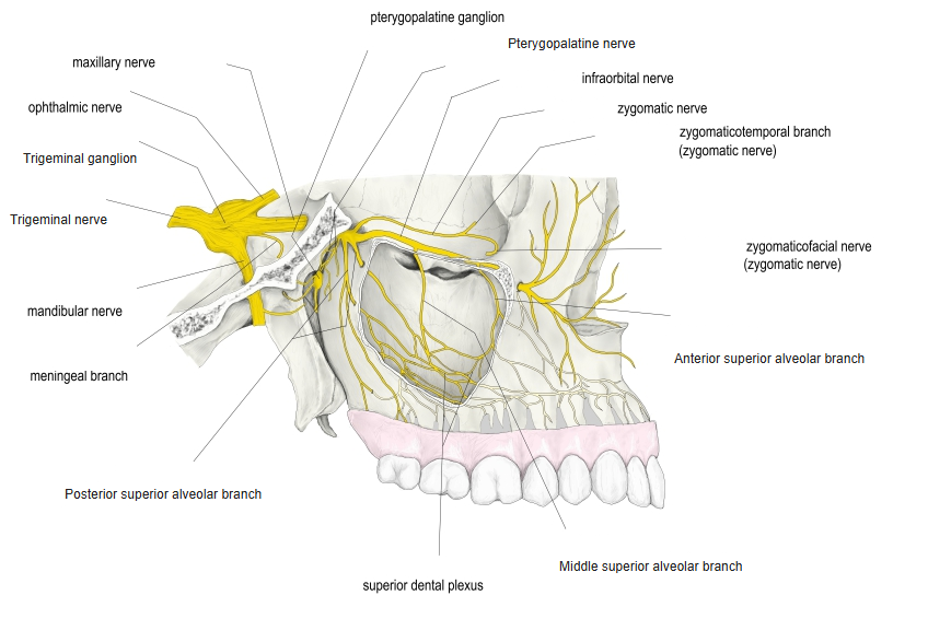
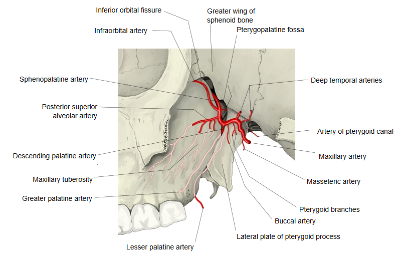
Nerves and vessels: the posterior superior alveolar branches (of the maxillary nerve), the posterior superior alveolar artery and the posterior superior alveolar veins
· In shape it is roughly triangular.
· It forms where the back of the maxilla and the pterygoid process diverge from each other; it is widest above and closes below, where the two bones meet near the maxillary tuberosity.
· Above, it meets the medial end of the inferior orbital fissure at a right angle.
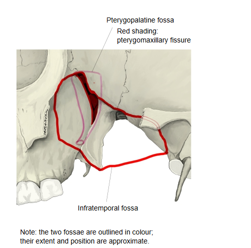
The pterygomaxillary fissure can be regarded as roughly the lateral side of the pterygopalatine fossa (a space shaped like a pyramid that narrows downwards).
Pterygopalatine fossa: a space medial to the infratemporal fossa that communicates with the nasal cavity and the orbit
· It is the lateral opening of the pterygopalatine fossa and connects it with the infratemporal fossa. The last part of the maxillary artery passes in through it; the posterior superior alveolar nerve, artery and veins pass out of it to the back of the maxilla.
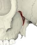 Pterygomaxillary fissure (lateral view)
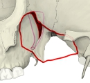 Infratemporal fossa and pterygopalatine fossa
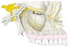 Maxillary nerve
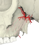 Maxillary artery in the pterygopalatine fossa
Reference websites
[Wikipedia]
· Pterygomaxillary fissure, Wikipedia.
Related terms
- Pterygopalatine fossa
- Infratemporal fossa
- Inferior orbital fissure
- Pterygoid process
- Maxillary tuberosity
- Maxillary artery
- Maxillary nerve
References and sources
- Further reading: Pterygomaxillary fissure, Wikipedia.
- Federative International Programme for Anatomical Terminology (FIPAT). Terminologia Anatomica, 2nd ed. (TA2), 2019.