Lymphatic trunk (Latin: truncus lymphaticus)
Also called the lymph trunk
Overview
From lymph capillaries to the veins
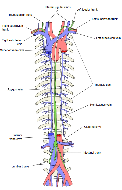
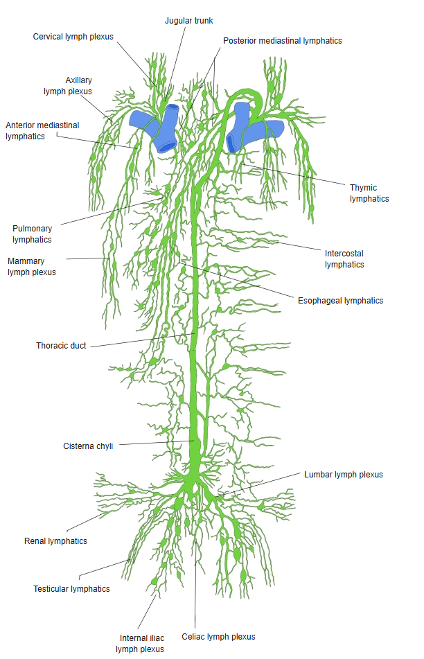
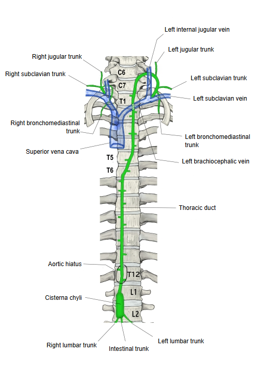
The thoracic duct is sometimes counted as a kind of lymphatic trunk. It begins with a dilatation, the cisterna chyli, and carries about three quarters of the body's lymph to the veins.
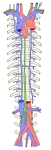 Lymphatic trunks
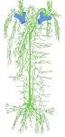 Lymph vessels of the trunk
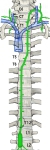 Lymphatic trunks, 2
Structure
The wall of a lymphatic trunk has the following three layers.
| Layer | Make-up | |
|---|---|---|
| 1 | Tunica intima | Endothelial cells on a thin basement membrane. It forms many valves that keep the lymph flowing towards the veins. |
| 2 | Tunica media | A few layers of smooth muscle fibres with some elastic fibres; their contractions push the lymph onward. |
| 3 | Tunica externa | Loose connective tissue that blends with the surrounding tissue; it may carry small blood vessels and nerves. |
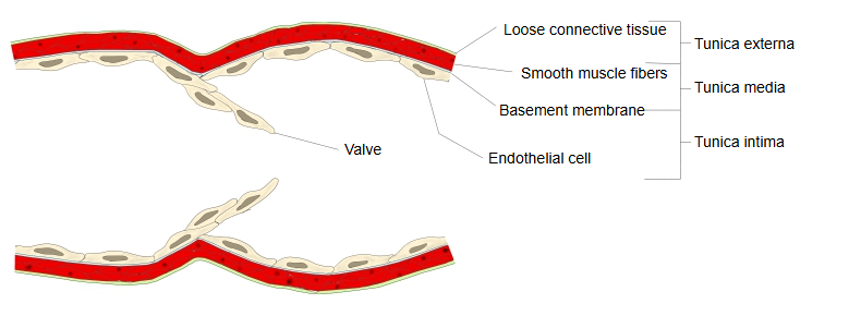
· Unlike arteries and veins, the three layers are thin and their boundaries are not clear.
· Illustration: collecting lymphatic vessel and lymphatic capillary (MDPI)

· Illustration: wall and valves of a lymph vessel (lymphangion) (Springer)

Types
| Name | Remarks | ||
|---|---|---|---|
| Right venous angle | 1 | Right jugular trunk | Collects the lymph of the head and neck of the right side |
| 2 | Right bronchomediastinal trunk | Collects the lymph of the right lung and bronchi, part of the heart and the right side of the mediastinum | |
| 3 | Right subclavian trunk | Collects the lymph of the right upper limb, part of the right chest wall and the right breast | |
| Left venous angle | 1 | Left jugular trunk | Collects the lymph of the head and neck of the left side |
| 2 | Left bronchomediastinal trunk | Collects the lymph of the left lung and bronchi, part of the heart and the left side of the mediastinum | |
| 3 | Left subclavian trunk | Collects the lymph of the left upper limb, part of the left chest wall and the left breast | |
| 4 | Lumbar trunks (right and left) | Collect the lymph of the lower limbs, the pelvic organs, the kidneys and other retroperitoneal organs, and the lower abdominal wall | |
| 5 | Intestinal trunk | Collects the lymph of the stomach, intestine, liver, pancreas and spleen; after a meal it is milky chyle |
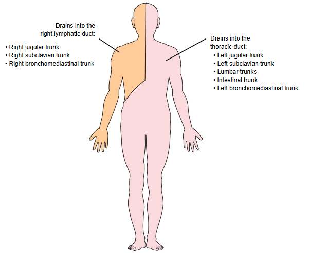
· Right side (head, neck and thorax of the right side, right upper limb): the right jugular, subclavian and bronchomediastinal trunks join the right lymphatic duct, or open one by one into the right venous angle.
· Left side: the left jugular, subclavian and bronchomediastinal trunks join the end of the thoracic duct, or open separately into the left venous angle.
· Abdomen: the lumbar and intestinal trunks meet at the cisterna chyli, the lower end of the thoracic duct.
· Sources differ on the pattern of the trunks: on either side they may join one another or open separately into the veins, and the intestinal trunk is often double or multiple.
Related lymph nodes
| Lymphatic trunk | Main related lymph nodes | Main areas drained | |
|---|---|---|---|
| 1 | Lumbar trunk | Lumbar lymph nodes (which also receive the iliac lymph nodes) | Lower limb, pelvic organs |
| 2 | Intestinal trunk | Coeliac and mesenteric lymph nodes | Gastrointestinal tract, liver, spleen |
| 3 | Bronchomediastinal trunk | Tracheobronchial and mediastinal lymph nodes | Lungs, trachea, heart |
| 4 | Subclavian trunk | Axillary (apical) lymph nodes | Upper limb, mammary gland |
| 5 | Jugular trunk | Deep cervical lymph nodes | Head, face, neck |
Reference websites
[Wikipedia]
· Lymphatic vessel, Wikipedia.
· Thoracic duct, Wikipedia.
Related terms
- Subclavian trunk
- Thoracic duct
- Right lymphatic duct
- Cisterna chyli
- Jugular trunk
- Venous angle
- Lymph node
- Lymphatic plexus
References and sources
- Further reading: Lymphatic vessel, Wikipedia.
- Further reading: Thoracic duct, Wikipedia.
- Federative International Programme for Anatomical Terminology (FIPAT). Terminologia Anatomica, 2nd ed. (TA2), 2019.