Shoulder joint (Latin: articulatio humeri; articulatio glenohumeralis)
Also called the glenohumeral joint
What is the shoulder joint?
- It is a typical multiaxial ball-and-socket joint.
- It has the widest range of movement of any joint in the body.
- The socket (glenoid cavity) is small compared with the head (head of the humerus): it covers only about a third of the articular surface of the head. A rim of fibrocartilage, the glenoid labrum, deepens it.
- It is also called the glenohumeral joint.
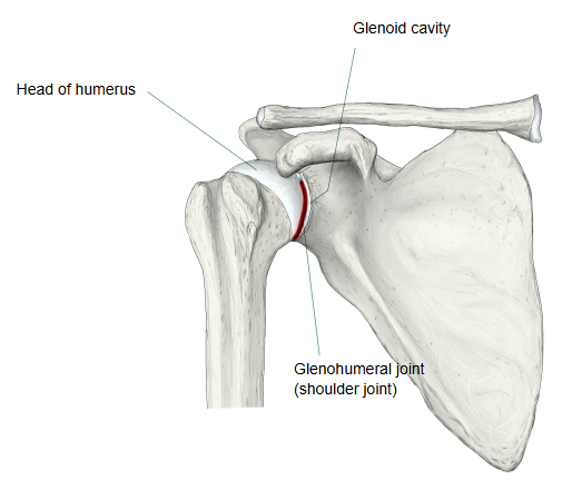
Bones
Type
Ligaments
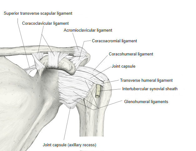
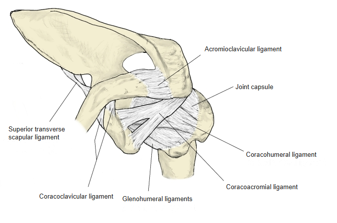
Three ligaments belong to the shoulder joint itself. The first two reinforce the joint capsule.
| Ligament | Course | |
|---|---|---|
| 1 | Coracohumeral ligament | From the base of the coracoid process to the greater and lesser tubercles of the humerus, blending with the upper part of the capsule. |
| 2 | Glenohumeral ligaments (superior, middle, inferior) | Thickenings of the front of the capsule, from the margin of the glenoid cavity and glenoid labrum to the anatomical neck and lesser tubercle of the humerus. |
| 3 | Transverse humeral ligament | Bridges the intertubercular groove between the greater and lesser tubercles, holding the tendon of the long head of biceps brachii in the groove. |
Besides these three, the Coracoacromial ligament is often listed with the shoulder joint: it runs from the coracoid process to the acromion and forms, with them, the coraco-acromial arch over the head of the humerus.
Movements / muscles
The shoulder joint has the widest range of movement of all the joints in the body. Its movements are enlarged by the joints of the pectoral girdle (sternoclavicular and acromioclavicular joints), which move the scapula at the same time. The glenoid cavity does not face straight laterally: it faces forwards and laterally, in a plane about 30° in front of the frontal plane (the plane of the scapula). Movement of the arm is freest in this plane, which is also where the arm usually works, for example when writing or eating.
| Movement | Description | Main muscles | |
|---|---|---|---|
| 1 | Flexion | The arm moves forwards and upwards in the sagittal plane. | Deltoid (anterior part) Pectoralis major Coracobrachialis |
| 2 | Extension | The arm moves backwards in the sagittal plane. | Latissimus dorsi Deltoid (posterior part) |
| 3 | Adduction | The arm moves towards the trunk in the frontal plane. | Pectoralis major Latissimus dorsi Teres major Subscapularis |
| 4 | Abduction | The arm moves away from the trunk in the frontal plane. | Deltoid Supraspinatus Trapezius (see the note) Serratus anterior (see the note) |
| 5 | Medial (internal) rotation | The arm turns inwards about its long axis. | Deltoid (anterior part) Teres major Latissimus dorsi Subscapularis Pectoralis major |
| 6 | Lateral (external) rotation | The arm turns outwards about its long axis. | Deltoid (posterior part) Infraspinatus Teres minor |
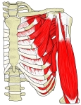
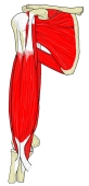
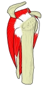
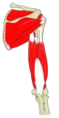
Note: the deltoid and the supraspinatus raise the arm sideways at the shoulder joint. Raising the arm further, above the shoulder, needs the scapula to rotate upwards as well: the Trapezius and the Serratus anterior do this, acting on the scapula rather than on the shoulder joint itself.
Other
Dislocation of the shoulder: the shoulder joint is the joint most often dislocated, about half of all dislocations. Because the socket is shallow and the capsule loose, the head of the humerus is easily forced out of the glenoid cavity, usually forwards and downwards. Typical causes are a blow or fall on the shoulder, or a backward force on the arm while it is raised.