Greater palatine canal (Latin: canalis palatinus major)
Also called the pterygopalatine canal
· Its length, from the pterygopalatine fossa to the greater palatine foramen, is about 3 cm on average, with wide variation between people.
Sources differ on which structures pass through the canal.
Some textbooks list the same vessels but name the nerve the "anterior palatine nerve", an older name for the greater palatine nerve.
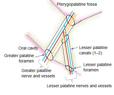
Wikipedia lists the descending palatine artery and vein and the greater and lesser palatine nerves; the lesser palatine nerves run in the upper part of the canal before entering the lesser palatine canals.
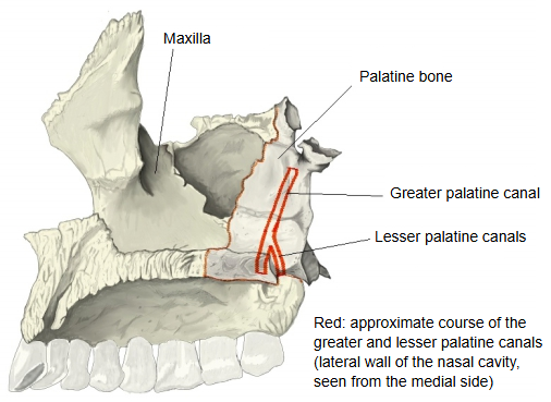
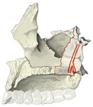 Lateral wall of the nasal cavity
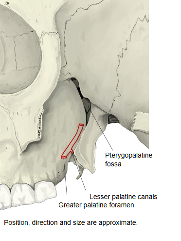
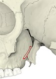 Skull (lateral, behind the maxilla)
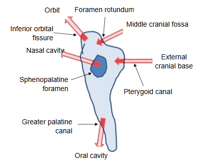
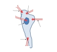 Pterygopalatine fossa (diagram)
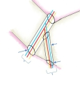 Greater and lesser palatine canals (diagram)
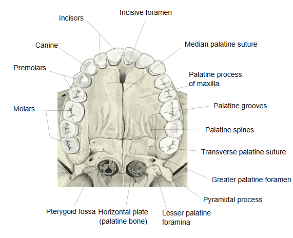
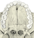 Hard palate (inferior)
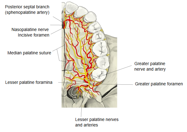
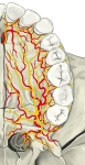 Nerves and arteries of the palate
Reference websites
[Wikipedia]
· Greater palatine canal, Wikipedia.
· Lesser palatine canals, Wikipedia.
[Websites with illustrations]
· The pterygopalatine ganglion and its branches (Gray's Anatomy, fig. 780) (Wikimedia Commons)

Structure
The canal begins at the lower end of the pterygopalatine fossa and passes between the maxilla and the palatine bone to the hard palate, ending at the greater palatine foramen at the back corner of the palate, near the lateral border of the horizontal plate. It is formed by the vertical greater palatine groove on the maxillary surface of the perpendicular plate of the palatine bone, which is closed into a canal by a matching groove on the maxilla. Accessory canals, the lesser palatine canals, branch off it. It transmits the descending palatine artery and vein, the greater palatine nerve and, in its upper part, the lesser palatine nerves. The greater palatine nerve can be blocked at the greater palatine foramen for dental work on the palate.
Where it is found
Related terms
- Perpendicular plate
- Horizontal plate
- Pyramidal process
- Hard palate
- Sphenopalatine foramen
- Palatine process
- Palatine bone
References and sources
- Further reading: Greater palatine canal, Wikipedia.
- Further reading: Pterygopalatine fossa, Wikipedia.
- Federative International Programme for Anatomical Terminology (FIPAT). Terminologia Anatomica, 2nd ed. (TA2), 2019.