Palatine process (Latin: processus palatinus maxillae)
- Body : the roughly pyramidal main part of the maxilla; it contains the maxillary sinus and has four surfaces.
- Processes
- Frontal process : a long, flat process that rises upward between the nasal bone and the lacrimal bone.
- Zygomatic process : a short, thick process from the lateral side of the body that joins the zygomatic bone.
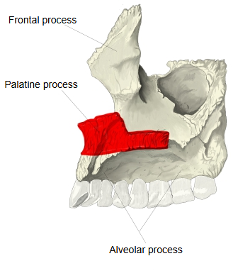
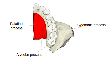
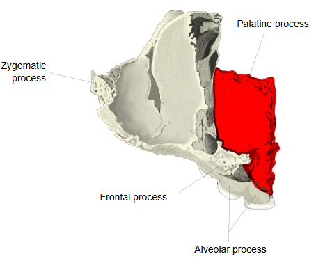
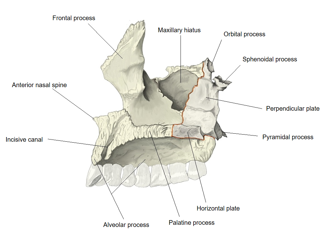
- Palatine process : projects almost horizontally inward from the lower part of the body.
- Alveolar process : the downward continuation of the body; it holds the tooth sockets for the roots of the upper teeth.
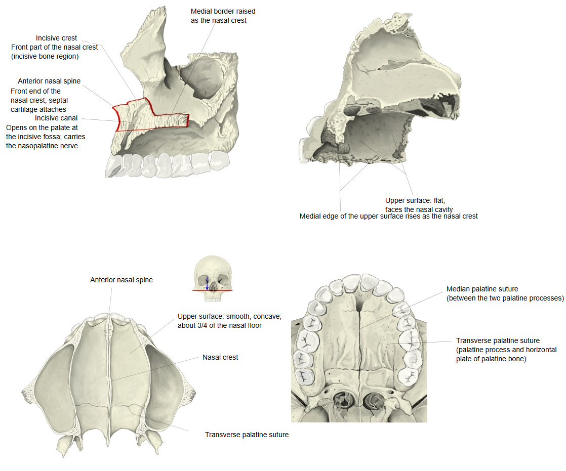
Upper (nasal) surface
· It is concave from side to side.
· It is smooth.
· It forms most of the floor of the nasal cavity.
· Near the front of its medial edge there is an opening that leads into the incisive canal.
Medial border
· The bone is thicker in front than behind.
· It rises into a ridge (the nasal crest), which meets the same ridge of the other maxilla and carries the vomer.
· The front part of the nasal crest is raised and is called the incisor crest.
· The front end of the incisor crest projects forward as the anterior nasal spine.
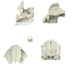 Palatine process (four views)
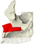 Right maxilla (medial)
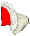 Right maxilla (inferior)
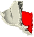 Right maxilla (superior)
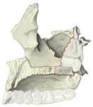 Maxilla and palatine bone (medial)
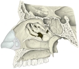 Lateral wall of nasal cavity
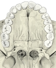 Hard palate (inferior)
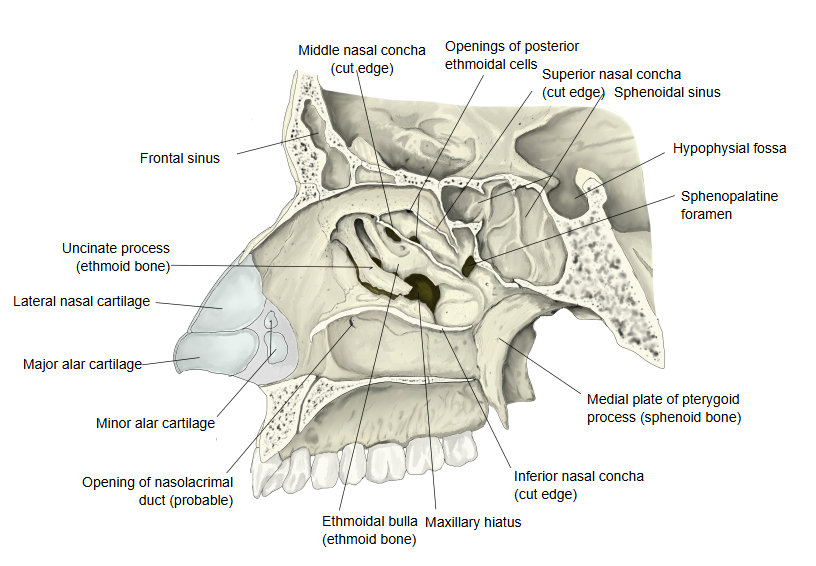
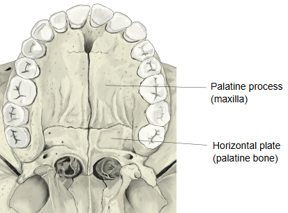
Lower (oral) surface
The lower surface is rough, with small pits for the palatine glands and many small foramina for nutrient vessels. Along the back part of its lateral border runs a groove (the palatine grooves), sometimes a canal, for the greater palatine nerve and vessels coming forward from the greater palatine foramen. The palatine processes of the two maxillae meet at the median palatine suture and form the front three quarters of the hard palate; behind, they meet the horizontal plates of the palatine bones at the transverse palatine suture. When the two maxillae are joined, the funnel-shaped incisive fossa is seen in the midline just behind the central incisors; the incisive canals open into it and transmit the nasopalatine nerve and the terminal branch of the greater palatine artery.
Note: in young skulls a faint incisive suture may be seen running from the incisive fossa toward the gap between the lateral incisor and the canine. It marks the boundary of the premaxilla (incisive bone), a separate bone in many animals.
Where it is found
Related terms
- Hard palate
- Horizontal plate
- Transverse palatine suture
- Incisive canal
- Anterior nasal spine
- Alveolar process
- Body of maxilla
References and sources
- Further reading: Palatine process of maxilla, Wikipedia.
- Further reading: Hard palate, Wikipedia.
- Federative International Programme for Anatomical Terminology (FIPAT). Terminologia Anatomica, 2nd ed. (TA2), 2019.