Body of mandible (Latin: corpus mandibulae)
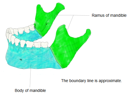
- Body of mandible : the flat, curved front part in which the roots of the lower teeth are set. Features: mental foramen, mental protuberance, mylohyoid line, alveolar part, etc.
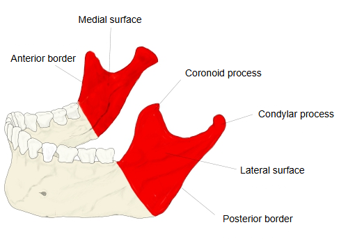
- Ramus of mandible : the paired back part, rising upwards and backwards on each side. Features: coronoid process, condylar process, mandibular notch, mandibular foramen, mylohyoid groove, etc.
Common to both parts: the angle of mandible and the mandibular canal.
The body is horseshoe-shaped. Its two halves meet in the midline at the mandibular symphysis and fuse during the first year of life. Besides the two surfaces and the upper alveolar part, it has a rounded lower border, the base of mandible. Behind, it continues on each side into the ramus.
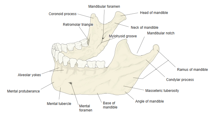
External surface: a faint midline ridge marks the symphysis. Below, it widens into the triangular mental protuberance (the chin), with a mental tubercle at each lower corner. The mental foramen opens on each side, usually below the second premolar, midway between the upper and lower borders; the mental nerve and vessels pass through it. The oblique line runs backwards and upwards from the mental tubercle and continues into the anterior border of the ramus. Muscles of the lower lip and chin (mentalis, depressor labii inferioris, depressor anguli oris) and the platysma attach here.
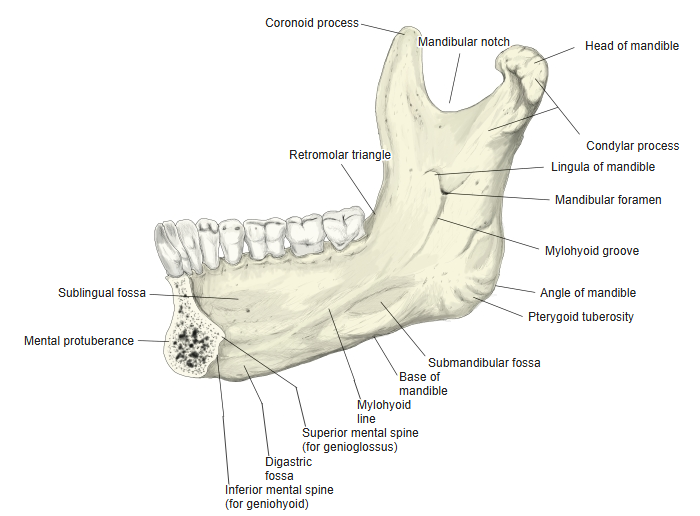
Internal surface: near the lower end of the symphysis are the mental spines: the superior pair for the genioglossus and the inferior pair for the geniohyoid. Below them, on each side of the midline, is the digastric fossa for the anterior belly of the digastric. The mylohyoid line runs upwards and backwards from near the symphysis; the mylohyoid arises from it. Above the front of the line is the sublingual fossa (sublingual gland); below its back part is the submandibular fossa (submandibular gland). Inside the body runs the mandibular canal, from the mandibular foramen on the ramus to the mental foramen.
Alveolar part and base: the alveolar part is hollowed into dental alveoli (tooth sockets), 16 in the full adult set. The roots of the front teeth raise the outer bone into alveolar yokes. The buccinator attaches along the outer side of the alveolar part in the molar region. After tooth loss the alveolar part is gradually resorbed, so the body becomes lower. The base of mandible is thick and rounded; near its junction with the ramus, the facial artery may leave a shallow groove as it crosses onto the face. The body meets the ramus at the angle of mandible.
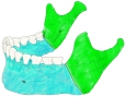 Body and rami of mandible
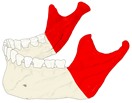 Ramus of mandible
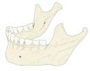 Mandible (oblique view)
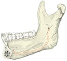 Right half of mandible (medial view)
Reference websites
[Wikipedia]
· Mandible, Wikipedia.
[Websites with illustrations]
· Mandible, outer surface, side view (Gray's Anatomy, fig. 176) (Wikimedia Commons)

· Mandible, inner surface, side view (Gray's Anatomy, fig. 177) (Wikimedia Commons)

· The mandible: structure and attachments (TeachMeAnatomy)
· The mandible: anatomy, structure, function (Kenhub)
Where it is found
Related terms
- Ramus of mandible
- Angle of mandible
- Mental foramen
- Mylohyoid line
- Mandibular canal
- Mandibular foramen
- Mandible
References and sources
- Further reading: Mandible, Wikipedia.
- Federative International Programme for Anatomical Terminology (FIPAT). Terminologia Anatomica, 2nd ed. (TA2), 2019.