Zygomatico-orbital foramen (Latin: foramen zygomaticoorbitale)
The zygomatic bone has three surfaces, as below; the zygomatico-orbital foramen lies on the one that faces the orbit, in the lateral wall of the orbit.
bone
- Lateral surface
- Orbital surface : carries the zygomatico-orbital foramen (one or two small openings), which transmits the zygomatic nerve.
- Temporal surface
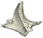 Zygomatic bone
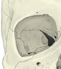 Right orbit (anterior view)
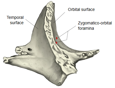
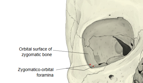
The foramen leads into small canals in the bone. One canal opens on the temporal surface at the zygomaticotemporal foramen, facing the temporal fossa; the other opens on the lateral (facial) surface at the zygomaticofacial foramen.
It transmits the zygomatic nerve (a branch of the maxillary nerve) or its two branches, the zygomaticotemporal and zygomaticofacial nerves, with small vessels; the first runs through the canal to the temporal surface, the second through the canal to the lateral surface.
There are usually one or two foramina. Sources differ: some describe a single foramen whose canal divides, others two separate foramina.
Reference websites
[Wikipedia]
· Zygomatico-orbital foramina, Wikipedia.
· Zygomatic bone, Wikipedia.
[Websites with illustrations]
· Left zygomatic bone (Gray's Anatomy, fig. 165) (Wikimedia Commons)

· Zygomatic bone: surfaces, processes, functions (Kenhub)
Where it is found
Related terms
References and sources
- Further reading: Zygomatico-orbital foramen, Wikipedia.
- Further reading: Zygomatic bone, Wikipedia.
- Federative International Programme for Anatomical Terminology (FIPAT). Terminologia Anatomica, 2nd ed. (TA2), 2019.