Latin: glandula parotidea
Overview
- The largest salivary gland, in front of and below the ear; a purely serous gland.
- Produces about a quarter of resting saliva; its share rises to about half when salivation is stimulated.
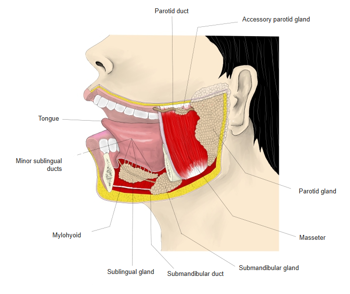
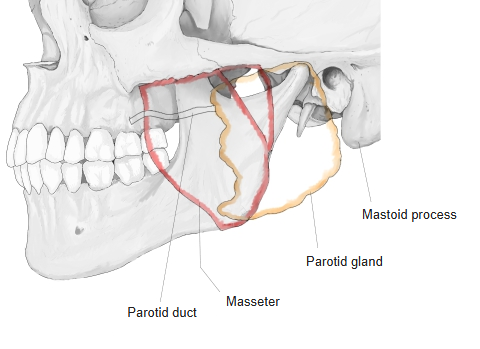
- Lies on the masseter and behind the ramus of the mandible, in the retromandibular fossa; enclosed in a tough parotid fascia.
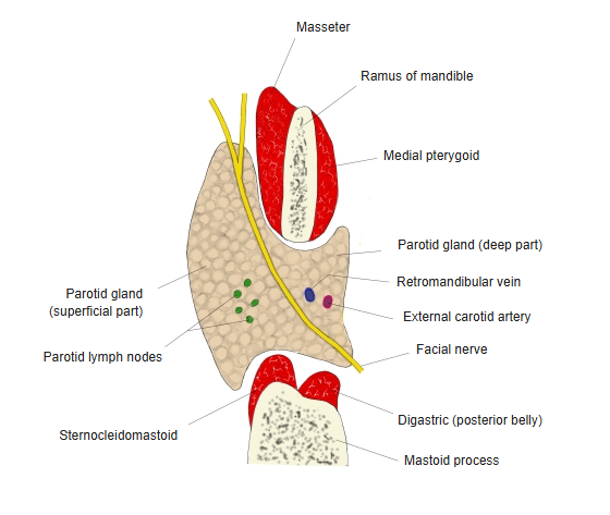
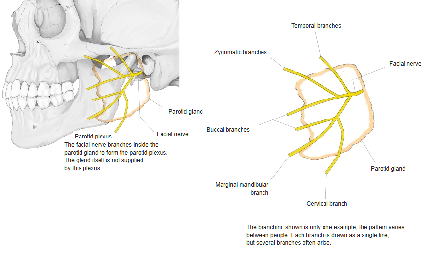
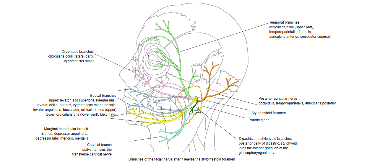
- The facial nerve passes through it and divides it into a large superficial part and a smaller deep part.
Major salivary glands 
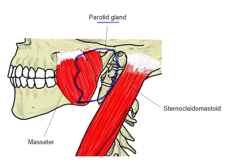
Parotid gland and masseter
Location and relations
- Superficial (lateral) surface: slightly convex, covered by skin, fascia and the parotid fascia.
- Anteromedial surface: against the masseter, ramus of the mandible and medial pterygoid.
- Posteromedial surface: against the mastoid process, sternocleidomastoid, posterior belly of the digastric and the styloid process.
- Upper border: zygomatic arch and external acoustic meatus; lower end: reaches the anterior border of the sternocleidomastoid.
- Structures inside the gland, from superficial to deep: facial nerve (parotid plexus), retromandibular vein, external carotid artery.
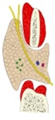
Parotid gland (horizontal section) 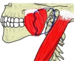
Parotid gland and neighbouring muscles 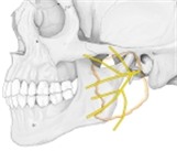
Parotid plexus
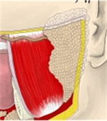
Parotid duct
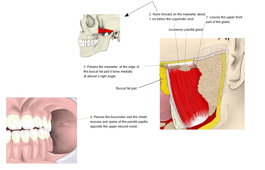
- Leaves the upper front part of the gland.
- Runs forward on the masseter about 1 cm below the zygomatic arch.
- At the front edge of the masseter and the buccal fat pad it turns medially almost at a right angle.
- Pierces the buccinator and the cheek mucosa and opens at the parotid papilla, opposite the upper second molar.
- An accessory parotid gland of variable size often lies along the duct and drains into it.
Course of the parotid duct
Blood supply, lymph and nerves
| Supply | Source |
|---|---|
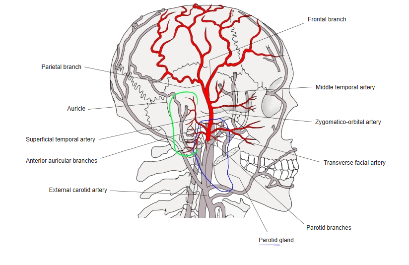
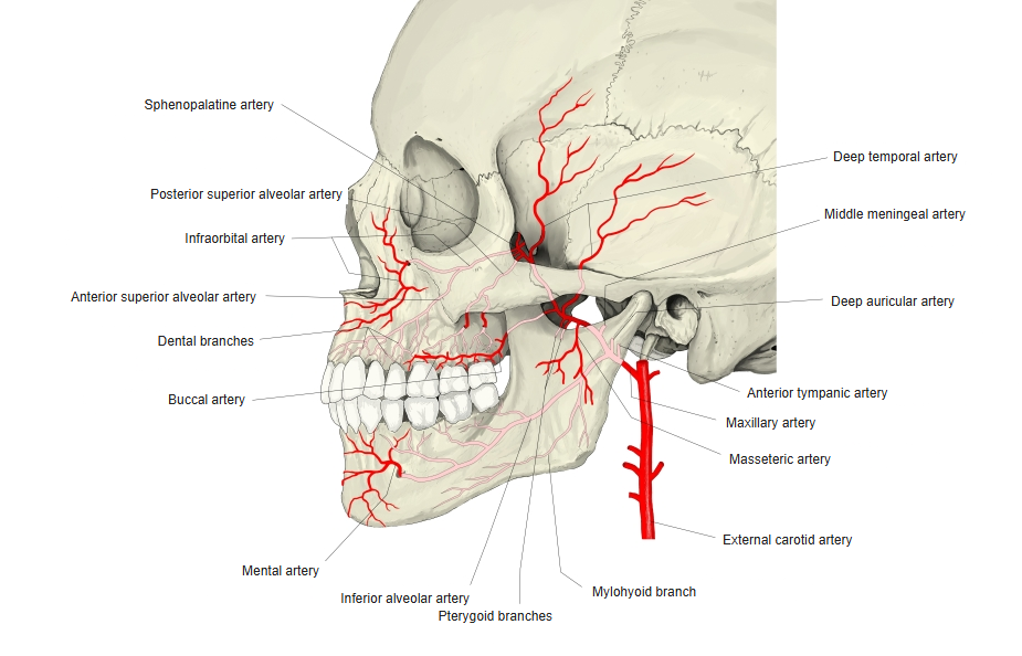
| Arteries | Branches of the external carotid artery in the gland: superficial temporal and maxillary arteries (including the transverse facial artery) |
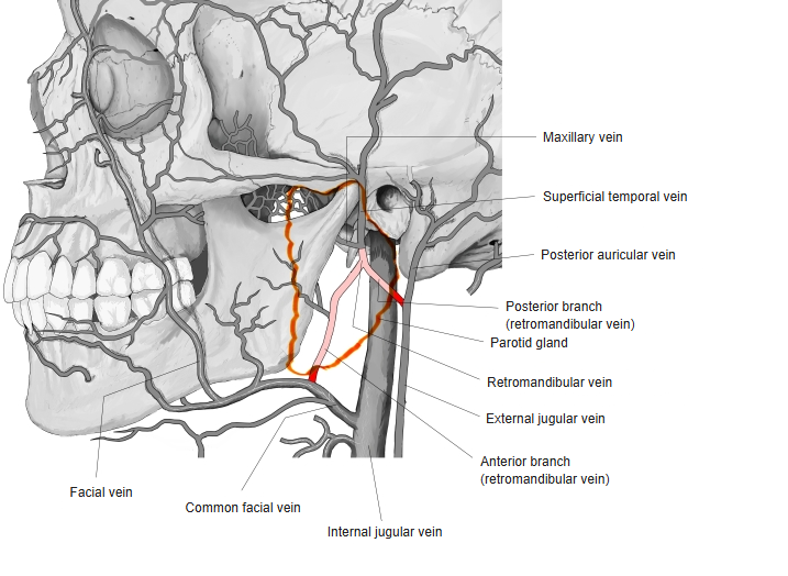
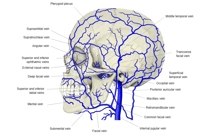
| Veins | Retromandibular vein; some to the facial vein |
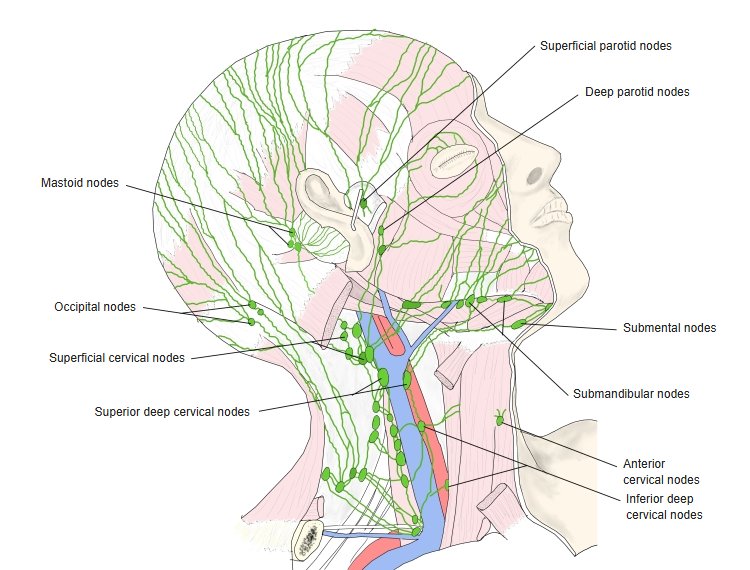
| Lymph | Superficial and deep parotid nodes, then the deep cervical nodes |
| Parasympathetic (secretomotor) | Glossopharyngeal nerve → lesser petrosal nerve → otic ganglion → auriculotemporal nerve |
| Sympathetic | Superior cervical ganglion, via the plexus around the external carotid artery (mainly vasoconstriction) |
| Sensory | Great auricular nerve (gland, fascia and overlying skin) and auriculotemporal nerve |
- The facial nerve only passes through the gland (parotid plexus); it does not supply it.
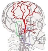
Superficial temporal artery 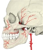
Maxillary artery 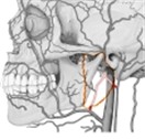
Retromandibular vein 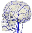
Veins of the head 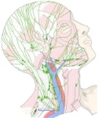
Lymph nodes of the head and neck 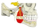
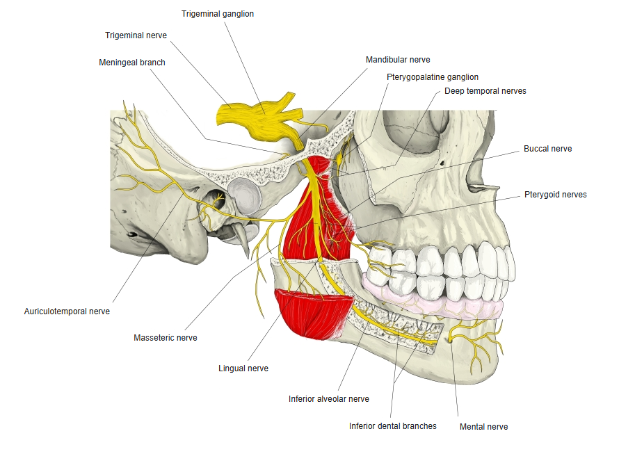
Mandibular nerve 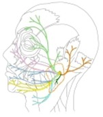
Facial nerve branches
References and sources
- Further reading: Parotid gland, Wikipedia.