Latin: structurae oculi accessoriae
Overview
- The parts of the visual organ other than the eyeball and optic nerve that protect, moisten and move the eyeball.
- Also called the accessory organs of the eye or ocular adnexa.
- What is included varies between books: some add the orbital fasciae and fat body, or even the bony orbit.
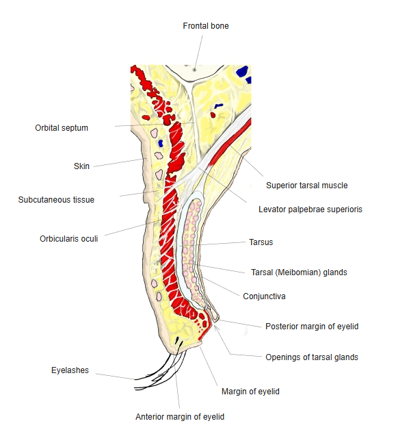
Upper eyelid, sagittal section 
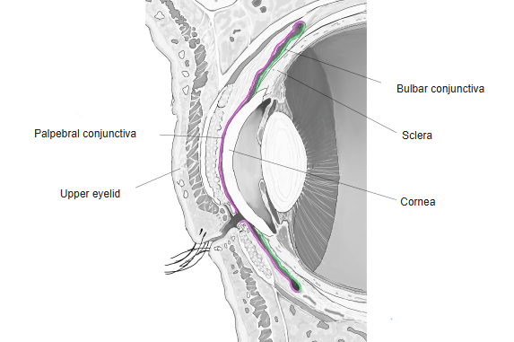
Conjunctiva, sagittal section
Accessory structures
| Structure | Role |
|---|---|
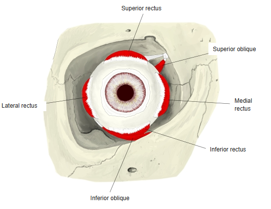
| Extraocular muscles | Six muscles that move the eyeball: superior, inferior, medial and lateral rectus, superior and inferior oblique; the levator palpebrae superioris raises the upper eyelid |
| Eyelids | Cover and protect the front of the eye; blinking spreads the tear film; eyelashes grow from their margins |
| Conjunctiva | Thin mucous membrane lining the back of the eyelids and covering the white of the eye |
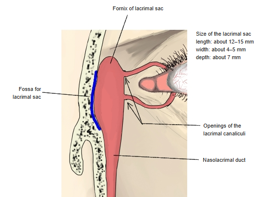
| Lacrimal apparatus | The lacrimal gland makes tears; the lacrimal canaliculi, lacrimal sac and nasolacrimal duct drain them into the nose |
| Eyebrows | Keep sweat from running into the eyes |
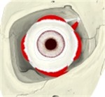
Right extraocular muscles, anterior view 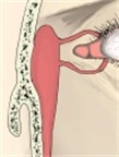
Lacrimal sac
References and sources
- Further reading: Accessory structures of eye, Wikipedia.