Adductor canal (Latin: canalis adductorius)
Also called: subsartorial canal; Hunter's canal
· It is also called Hunter's canal, or the subsartorial canal.
Some texts divide the adductor canal into an upper muscular part and a lower tendinous part.
canal
- Muscular part : the upper part, bounded by adductor longus, vastus medialis and sartorius
- Tendinous part : the lower part, covered by the vastoadductor membrane
 Right thigh muscles (anterior)
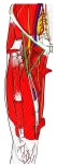 Femoral triangle and adductor canal
- Muscular and tendinous parts of the adductor canal
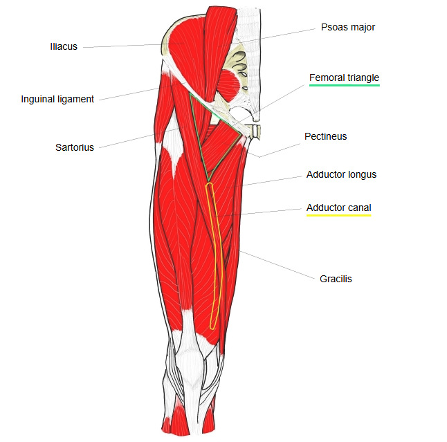
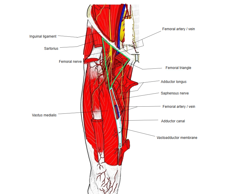
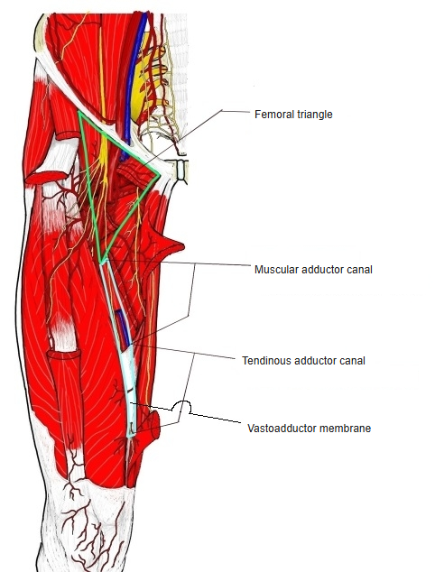
Below the iliopectineal fossa lies the muscular part of the canal, bounded by adductor longus, vastus medialis and sartorius; below it follows the tendinous part. Where adductor magnus inserts on the linea aspera of the femur, a long aponeurotic sheet (the vastoadductor membrane) runs from its front surface to the medial side of vastus medialis; the tendinous part is the channel between these two muscles under that sheet. At its lower end the insertion tendon of adductor magnus forms a tendinous arch, leaving the adductor hiatus between it and the femur. The femoral artery and vein run from the iliopectineal fossa through the adductor canal and the hiatus to the back of the knee.
Reference websites
[Wikipedia]
· Adductor canal, Wikipedia.
· Adductor hiatus, Wikipedia.
[Websites with illustrations]
· The adductor canal: borders and contents (TeachMeAnatomy)
· Contents of the adductor canal (Earth's Lab)
Structure
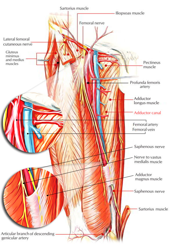
The adductor canal is a narrow intermuscular tunnel on the medial side of the middle third of the thigh, running from the apex of the femoral triangle to the adductor hiatus. Its walls are:
Anteromedially: a tough fibrous roof, the vastoadductor membrane, with sartorius lying on it.
Posteriorly: adductor longus and adductor magnus.
Laterally: vastus medialis.
The roof stretches from vastus medialis across the femoral vessels to adductor longus and adductor magnus.
Sartorius lies on this roof.
Contents: the femoral artery and femoral vein, and two branches of the femoral nerve: the saphenous nerve and the nerve to vastus medialis. The vessels and the saphenous nerve enter at the upper end. The saphenous nerve does not pass the hiatus: with the saphenous branch of the descending genicular artery it pierces the roof of the canal and runs on to the leg. The femoral vessels leave at the lower end through the adductor hiatus, a gap in the adductor magnus tendon, and become the popliteal artery and popliteal vein behind the knee. The saphenous nerve is blocked here with local anaesthetic (adductor canal block) for pain relief after knee surgery.
History
The name Hunter's canal honours the Scottish surgeon John Hunter.
Where it is found
Related terms
- Femoral triangle
- Sartorius
- Vastus medialis
- Adductor magnus
- Femoral artery
- Saphenous nerve
- Femoral vein
References and sources
- Further reading: Adductor canal, Wikipedia.
- Further reading: Adductor hiatus, Wikipedia.
- Federative International Programme for Anatomical Terminology (FIPAT). Terminologia Anatomica, 2nd ed. (TA2), 2019.