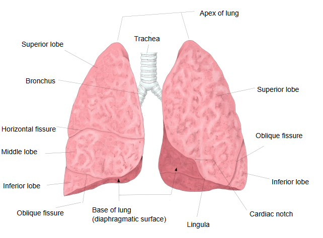
Apex of lung (Latin: apex pulmonis)
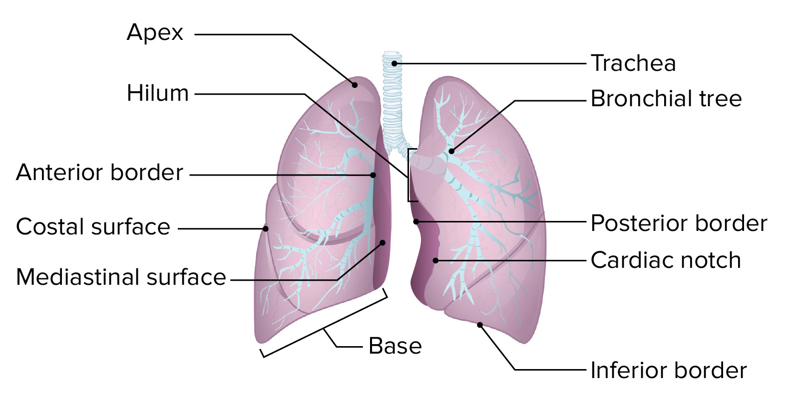
- Apex : the blunt, rounded top, rising about 2–3 cm above the clavicle and covered by the cervical pleura. Grooves: for the subclavian artery (both lungs), for the left brachiocephalic vein (left) and for the superior vena cava (right).
- Three surfaces : the costal surface (the largest, facing the ribs), the medial surface (where the bronchus, vessels and nerves enter and leave) and the diaphragmatic surface (= the base).
- Three borders : the anterior border (between the costal and medial surfaces in front), the inferior border (between the costal and diaphragmatic surfaces) and the posterior border (between the costal and medial surfaces behind).
- Base : concave, following the dome of the diaphragm; = the diaphragmatic surface.
The apex shows grooves made by arteries and veins.
the apex
- Right lung : groove for the subclavian artery; groove for the superior vena cava.
- Left lung : groove for the subclavian artery; groove for the left brachiocephalic vein.
The shape and relations of the apex in more detail:
The apex is the blunt, rounded upper end of the lung. In front it projects about 2–3 cm above the upper border of the clavicle, over its medial third; behind it does not rise above the neck of the 1st rib, because the rib slopes downward toward the front. It is covered by the cervical pleura (pleural cupula), which is strengthened by the suprapleural membrane.
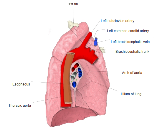
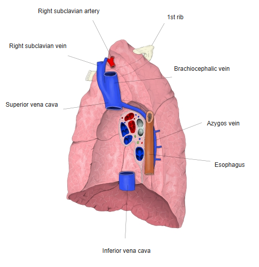
On the medial side of the apex of both lungs there is a groove for the subclavian artery, the groove for subclavian artery.
In front of it, the left lung has a groove for the left brachiocephalic vein, made by the left brachiocephalic vein, and the right lung has grooves for the right brachiocephalic vein and the superior vena cava (the groove for the superior vena cava).
Behind the apex lie the neck of the 1st rib, the sympathetic trunk and the anterior rami of the 8th cervical and 1st thoracic spinal nerves (C8 and T1).
A tumour of the apex (Pancoast tumour) can invade these nerves, causing pain in the arm and Horner syndrome. A needle inserted above the clavicle can puncture the apex and cause a pneumothorax.
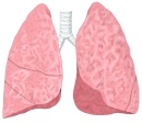 Lungs (anterior view)
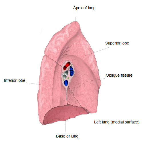
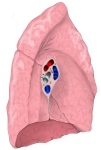 Left lung (medial surface)
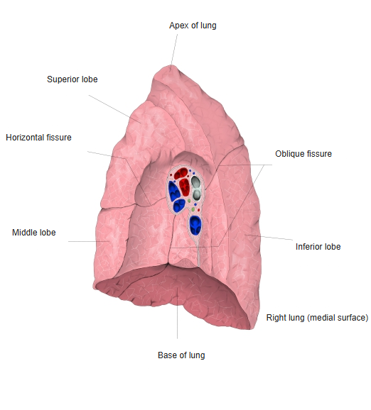
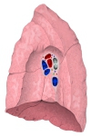 Right lung (medial surface)
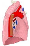 Left lung with nearby vessels (medial view)
 Right lung with nearby vessels (medial view)
Reference websites
[Wikipedia]
· Lung, Wikipedia.
· Pancoast tumor, Wikipedia.
[Websites with illustrations]
· The basic structure of the lung (figure) (Lecturio)
· The lungs (TeachMeAnatomy)
Where it is found
Related terms
References and sources
- Further reading: Lung, Wikipedia.
- Further reading: Pancoast tumor, Wikipedia.
- Federative International Programme for Anatomical Terminology (FIPAT). Terminologia Anatomica, 2nd ed. (TA2), 2019.