Serous membrane (Latin: tunica serosa)
Also called the serosa
- Overview
- Where it is found
- Structure
- Thickness
- Serosa, adventitia and capsule compared
- Reference websites
- (Wikipedia, websites with illustrations)
- Related terms
Overview
Together with the serous pericardium around the heart and the tunica vaginalis around the testis, these are the serous membranes of the body.
membrane
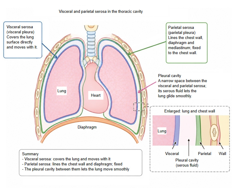
- Visceral layer : closely attached to the surface of an organ and follows its movements; e.g. the visceral peritoneum, the visceral pleura.
- Parietal layer : lines the inside of the body cavity; rich in sensory nerves, fairly thin and even; e.g. the parietal peritoneum, the parietal pleura.
Between the two layers is a narrow, closed space, such as the pleural cavity or the peritoneal cavity, holding a thin film of serous fluid that lets organs such as the lungs, heart and intestines move with little friction.
Where it is found
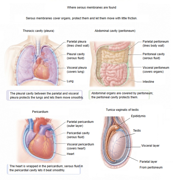
The table below lists where serous membranes are found, by the name of each membrane.
| Membrane | Main sites and organs | Role and features | |
|---|---|---|---|
| 1 | Peritoneum | Stomach, small intestine, large intestine (not all of it), liver, spleen, inner surface of the abdominal wall | Lines the abdominal and pelvic walls and covers the surfaces of the organs there. |
| 2 | Pleura | Lungs, inner surface of the chest wall (thorax), diaphragm and mediastinum | Wraps each lung and reduces the friction of breathing. |
| 3 | Serous pericardium | Heart | Covers the heart and lines the fibrous pericardium; protects and lubricates the beating heart. |
| 4 | Tunica vaginalis | Testis (the organ that makes sperm and testosterone) | Covers the front and sides of the testis; a pouch of peritoneum carried down into the scrotum when the testis descends. |
Structure
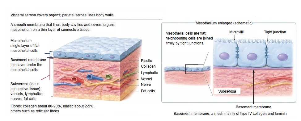
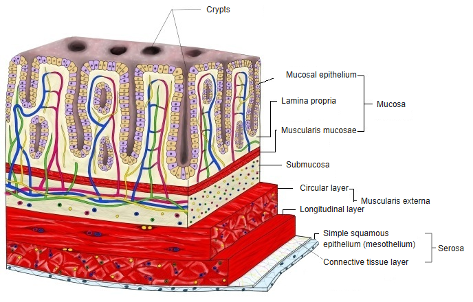
A serous membrane is made of a layer of flat cells one cell thick (the mesothelium) and the connective tissue layer beneath it.
· Basement membrane: a mesh-like sheet made mainly of type IV collagen and laminin (a glycoprotein).
Connective tissue layer
| Component | Description | Notes | |
|---|---|---|---|
| 1 | Collagen fibres | Types I and III, loosely arranged | Structural support; strength together with flexibility; mainly type I |
| 2 | Elastic fibres | A small amount | Let the membrane stretch and follow the movements of the organ |
| 3 | Fibroblasts | Connective tissue cells | Make and repair the fibres |
| 4 | Capillaries | A fine network of vessels | Supply the mesothelium and the surface tissue |
| 5 | Lymphatic vessels | Thin tubes | Take up serous fluid and cells |
| 6 | Nerve fibres | Fine branches of autonomic nerves | Control of the vessels; sensation |
| 7 | Fat cells | More in some places than in others | Cushioning and protection (e.g. the fatty appendices of the colon) |
| 8 | Immune cells | Macrophages and others | Local defence; inflammation |
The serosa is joined to the organs or to the abdominal and pelvic walls by the subserous tissue (subserosa), a layer of loose connective tissue that contains fat and elastic fibres; its thickness varies from place to place. In the subserous tissue of the greater omentum there are small clusters of immune cells known as milky spots.
Cell layer
| Component | Description | Role | |
|---|---|---|---|
| 1 | Mesothelial cells | Simple squamous epithelium | Form the outermost surface of the serosa |
| 2 | Microvilli | Fine projections of the cell surface | Hold serous fluid; reduce friction |
| 3 | Cell junctions | Tight junctions and others | Form a barrier; keep the layer stable |
| 4 | Basement membrane | Just beneath the mesothelium | Supports the cells; the base for exchange of substances |
Thickness
· Mesothelium: about 1–3 µm; loose connective tissue: about 5–50 µm; total: about 10–60 µm. These are rough values; sources differ.
| Site | Thickness | Features | |
|---|---|---|---|
| 1 | Jejunum and ileum | about 10–20 µm | Very thin and flexible |
| 2 | Stomach | about 20–40 µm | Somewhat thicker |
| 3 | Transverse colon | about 15–30 µm | Medium |
| 4 | Sigmoid colon | about 20–40 µm | Fairly thick |
| 5 | Caecum | about 15–25 µm | Rather thin |
| 6 | Spleen | about 30–60 µm | Among the thickest |
| 7 | Surface of the liver | about 20–50 µm | Fused with the capsule |
| 8 | Gallbladder | about 10–20 µm | Very thin |
Serosa, adventitia and capsule compared
around organs
and tubes
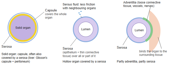
- Serosa : structure: a simple squamous epithelium (mesothelium) on a thin layer of connective tissue; secretes serous fluid that reduces friction. It covers organs that face a body cavity: the pleura, pericardium and peritoneum (each organ's part has its own name), e.g. the small and large intestines (in part).
- Adventitia : structure: loose connective tissue with vessels and nerves and no epithelium; anchors the organ to the surrounding tissue, with no clear boundary. E.g. oesophagus, much of the duodenum, parts of the large intestine, large blood vessels, trachea and bronchi.
- Capsule : dense connective tissue that is part of a solid organ itself, so not the same as an "organ + membrane" arrangement; it can be covered further by a serosa or an adventitia.
- Not covered by these
- Open to the outside : oral cavity, nasal cavity, pharynx, larynx, external acoustic meatus, anal canal: covered by mucosa or skin.
- Central nervous system : brain and spinal cord: covered by the meninges.
- Others : bone: periosteum; muscle: fascia; skin: epidermis and dermis.
Capsule: a membrane (dense connective tissue) that is part of a solid organ; it may be covered further by a serosa or an adventitia.
Serosa: "cell layer + connective tissue layer"; covers organs facing a body cavity and secretes serous fluid that reduces friction.
Adventitia: "loose connective tissue + vessels and nerves"; anchors the organ to the surrounding tissue.
Reference websites
[Wikipedia]
· Serous membrane, Wikipedia.
· Peritoneum, Wikipedia.
· Pleural cavity, Wikipedia.
[Websites with illustrations]
· Types of tissues (tissue membranes) (OpenStax Anatomy and Physiology 2e)
· The peritoneum (TeachMeAnatomy)
Related terms
- Mesothelium
- Pleural cavity
- Peritoneal cavity
- Visceral peritoneum
- Parietal peritoneum
- Visceral pleura
- Tunica adventitia
References and sources
- Further reading: Serous membrane, Wikipedia.
- Federative International Programme for Anatomical Terminology (FIPAT). Terminologia Anatomica, 2nd ed. (TA2), 2019.