Peritoneal folds (Latin: plicae peritoneales)
Also called Umbilical folds (folds of the anterior abdominal wall)
Overview
Peritoneal folds are fold-like ridges of the peritoneum that covers the inside of the abdominal cavity, raised along vessels or cords running beneath it.
= parietal peritoneum (a kind of serous membrane)
· Unlike a mesentery, a fold does not suspend an organ; it is more like a crease or wrinkle in the peritoneum lining the wall.
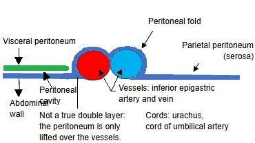
· Peritoneal folds of the abdominal wall are one of the structures of the peritoneum that reach into the peritoneal cavity. In a wider sense, "peritoneal folds" can mean any fold of peritoneum, including the folds around the duodenum, caecum and bladder; this page deals with the folds of the anterior abdominal wall.
Structures
of the
peritoneum
reaching into
the abdominal
cavity
of the
peritoneum
reaching into
the abdominal
cavity
- Mesentery : a double layer of peritoneum that joins an organ to the parietal peritoneum of the wall; the path of its vessels and nerves. Examples: the mesentery of the small intestine, the transverse mesocolon and others.
- Peritoneal folds : mostly a single layer: ridges on the inner surface of the anterior abdominal wall raised by vessels and cords. Examples: the median, medial and lateral umbilical folds.
- Omentum : peritoneum that wraps the stomach and continues up and down from its borders. Lesser omentum: stomach to liver, a double layer. Greater omentum: hangs down like a curtain; the double layer is folded back on itself, so its central part is a fused four-layered sheet.
- Peritoneal ligaments : a double layer that joins an organ to the abdominal wall or to another organ and holds it in place. Examples: the falciform ligament, the gastrosplenic ligament.
Types
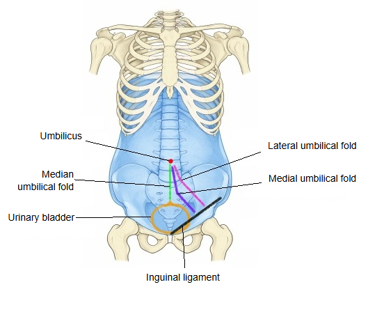
There are three peritoneal folds of the anterior abdominal wall: the median umbilical fold, the medial umbilical fold and the lateral umbilical fold, but only the lateral umbilical fold contains structures that still work.
Median and medial folds: their contents are fetal remnants without functionLateral fold: its contents are working blood vessels
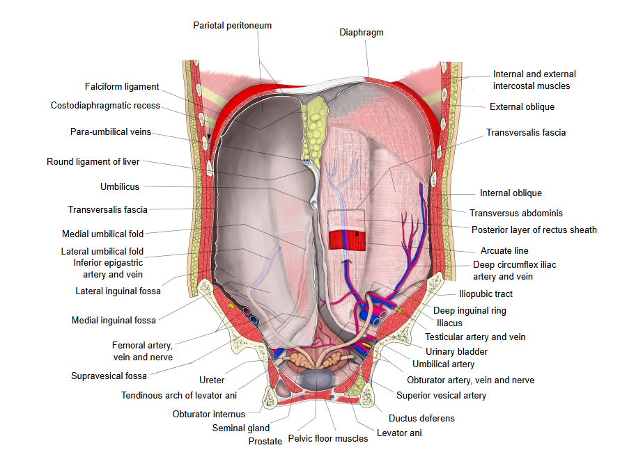
Folds and fossae of the anterior wall of the peritoneum
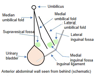
- Median umbilical fold : single, in the midline; joins the umbilicus and the apex of the urinary bladder. It covers the median umbilical ligament, the remnant of the fetal urachus.
- Supravesical fossa : between the lower parts of the median and medial umbilical folds, above the bladder.
- Medial umbilical fold : paired, lateral to the median umbilical fold. It covers the cord of the umbilical artery (medial umbilical ligament), the closed remnant of the fetal umbilical artery.
- Medial inguinal fossa : between the lower parts of the medial and lateral umbilical folds; site of a direct inguinal hernia.
- Lateral umbilical fold : paired, lateral to the medial umbilical fold. It contains the working inferior epigastric artery and vein.
- Lateral inguinal fossa : between the lower part of the lateral umbilical fold and the inguinal ligament, over the deep inguinal ring (entrance of the inguinal canal); site of an indirect inguinal hernia.
- Inguinal ligament : the band stretched between the pubic tubercle and the anterior superior iliac spine.
- Fold of the ductus deferens / round ligament of the uterus : formed by the ductus deferens (or the round ligament of the uterus), which passes from the deep inguinal ring, at the lower end of the lateral umbilical fold, medially and backwards into the pelvic cavity.
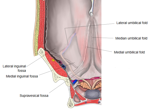
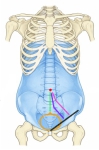 Position of the umbilical folds
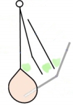 Folds and fossae (schematic)
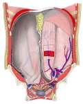 Anterior abdominal wall seen from behind
Related terms
- Median umbilical ligament
- Parietal peritoneum
- Peritoneal cavity
- Peritoneum
- Inferior epigastric artery
- Urinary bladder
- Inguinal ligament
References and sources
- Further reading: Median umbilical fold, Wikipedia.
- Further reading: Medial umbilical fold, Wikipedia.
- Further reading: Lateral umbilical fold, Wikipedia.
- Federative International Programme for Anatomical Terminology (FIPAT). Terminologia Anatomica, 2nd ed. (TA2), 2019.