Peritoneal ligaments
Also called Peritoneal ligament
Overview
- Mesentery : a double layer of peritoneum that attaches an organ to the parietal peritoneum of the abdominal wall and carries its blood vessels and nerves. Examples: the mesentery of the small intestine, the transverse mesocolon and others
- Peritoneal fold : a single layer raised into a ridge on the inner surface of the anterior abdominal wall by vessels or cord-like remnants. Examples: the median, medial and lateral umbilical folds
- Omentum : the peritoneum covering the stomach, extended from its borders upwards and downwards. Types: the lesser omentum (stomach to liver, a double layer) and the greater omentum (hangs down like a curtain; the double layer folds back on itself, so four layers fuse in the middle)
- Peritoneal ligament : a double layer that connects and supports an organ to the abdominal wall or to another organ. Examples: the falciform ligament, the gastrosplenic ligament
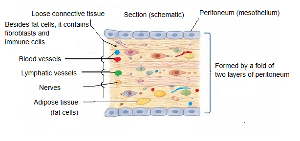
Although it is called a "ligament", a peritoneal ligament is quite different from the ligaments that join bones: it is a fold of serous membrane, not a band of dense fibrous tissue. Each one is two layers of peritoneum with a thin core of loose connective tissue, fat, small blood vessels, lymphatic vessels and nerves between them.
The table below sums up the differences.
| Feature | Peritoneal ligament | True ligament |
|---|---|---|
| Definition | A supporting structure of membrane, formed where the peritoneum folds double between organs or between organs and retroperitoneal structures | A supporting structure of fibrous connective tissue that joins bone to bone (or cartilage) directly |
| Tissue | Mesothelium (the surface cells of the peritoneum) plus loose connective tissue (mainly collagen fibres), with vessels, nerves and fat | Mainly dense regular connective tissue (rich in collagen bundles), fibroblasts and a few elastic fibres |
| Covering | Continuous with the peritoneum covering the organs (mesothelium on the surface) | No mesothelium; covered by a sheath of synovial membrane or connective tissue (or partly exposed inside a joint) |
| Vessels and nerves | Present (relatively many) | Few blood vessels, but many sensory and position-sense nerve endings |
| Surface | Smooth and moist (covered by serous fluid) | Dry fibrous surface |
| Flexibility | Soft and easily stretched | Strong; stretches very little |
| Main function | Holding organs in place, linking organs, and giving a path to vessels and nerves | Stabilising the skeleton; supporting and limiting the movement of joints |
| Examples | See Types below | The tibial collateral ligament, the anterior and posterior cruciate ligaments, the radial collateral ligament of the wrist and others |
Summary: a peritoneal ligament is a membranous structure formed by a fold of peritoneum; it contains many vessels and nerves and is soft and movable. A true ligament is made of dense fibrous connective tissue; it has few vessels and is a strong supporting tissue that hardly stretches.
Types
"Peritoneal ligament" is a general term, and sources differ on how many folds they count as peritoneal ligaments and on whether the same fold is called a ligament, a mesentery or part of an omentum (some texts write, for example, "falciform mesentery"). Most are named after the two structures they join, for example the gastrosplenic ligament (stomach and spleen).
(1) A short list
Here every fold is called a ligament.
| Ligaments that support the liver | ||
| 1 | Falciform ligament | The well-known sickle-shaped ligament joining the liver to the anterior abdominal wall (and the diaphragm); the round ligament of the liver runs in its free edge |
| 2 | Hepatogastric ligament | The membrane joining the liver and the stomach (part of the lesser omentum) |
| 3 | Coronary ligament / triangular ligaments | Ligaments that fix the liver closely to the diaphragm above it |
| Ligaments that support the stomach and spleen | ||
| 1 | Gastrocolic ligament | The large apron-like membrane joining the stomach and the transverse colon (part of the greater omentum) |
| 2 | Gastrosplenic ligament | Joins the stomach and the spleen |
| 3 | Splenorenal ligament | Joins the spleen to the posterior wall (in front of the left kidney) |
Ligament, mesentery and omentum
1. 1. Peritoneal ligaments: membranes that mainly fix organs in place; they carry few large vessels.
2. 2. Mesenteries: thick, important membranes that bring the large vessels and nerves, essential to life, to the organs (the small intestine and the large intestine), and attach the intestine to the posterior wall.
3. 3. Omenta (the lesser omentum and the greater omentum): large curtains of membrane centred on the stomach. When an omentum is divided into parts, the parts get names such as "hepatogastric ligament" or "gastrocolic ligament": the lesser omentum is made of the hepatogastric and hepatoduodenal ligaments, and the greater omentum includes the gastrocolic, gastrosplenic and gastrophrenic ligaments.
(2) A longer list
| Around the liver | ||
| 1 | Falciform ligament | Liver to the anterior abdominal wall and diaphragm |
| 2 | Coronary ligament | Liver to the diaphragm |
| 3 | Left triangular ligament | Left end of the coronary ligament, to the diaphragm |
| 4 | Right triangular ligament | Right end of the coronary ligament, to the diaphragm |
| 5 | Hepatogastric ligament | Liver to the lesser curvature of the stomach |
| 6 | Hepatoduodenal ligament | Liver to the duodenum |
| Around the stomach and spleen | ||
| 1 | Gastrophrenic ligament | Stomach to the diaphragm |
| 2 | Gastrosplenic ligament | Stomach to the spleen |
| 3 | Splenorenal ligament | Spleen to the posterior wall in front of the left kidney |
| 4 | Gastrocolic ligament | Stomach to the transverse colon |
| Around the colon | ||
| 1 | Phrenicocolic ligament | Left colic flexure to the diaphragm |
| Name | Main role |
|---|---|
| Peritoneal ligaments | Join one organ to another, or an organ to the abdominal wall or the diaphragm |
| Mesenteries | Suspend organs from the abdominal wall and give a path to vessels and nerves |
| Greater and lesser omentum | Special double folds of peritoneum that hang down from the stomach, or join the stomach and the liver |
Related terms
- Hepatogastric ligament
- Hepatoduodenal ligament
- Gastrocolic ligament
- Gastrosplenic ligament
- Splenorenal ligament
- Lesser omentum
- Mesentery
References and sources
- Further reading: Peritoneum, Wikipedia.
- Further reading: Ligament, Wikipedia.
- Federative International Programme for Anatomical Terminology (FIPAT). Terminologia Anatomica, 2nd ed. (TA2), 2019.