Tendon (Latin: tendo)
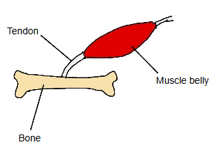
More details
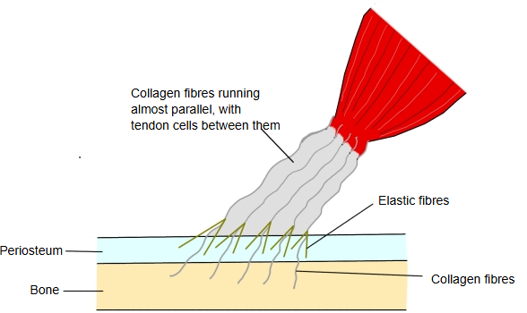
Tendon belongs to the dense connective tissue, one of the kinds of connective tissue. It is made mainly of collagen fibres (mostly type I) running parallel to the line of pull, with rows of tendon cells (tenocytes) between the fibres.
- Connective tissue proper
- 1. Loose connective tissue : holds organs and epithelia in place.
- 2. Dense connective tissue : forms ligaments and tendons.
- 3. Adipose tissue : made of fat cells.
- 4. Reticular tissue : supports the lymphoid organs.
- Special connective tissue : blood, bone and cartilage.
Connective tissue is also called supporting tissue.
Composition
Water makes up a large part of a living tendon; sources differ on the amount (figures from about 30% to 70% are quoted). Of the dry weight, the main components are:
| Component | Share of dry weight | |
|---|---|---|
| 1 | Collagen | most of it (roughly 65–85% in different sources) |
| 2 | Elastin (elastic fibres) | about 2% |
| 3 | Proteoglycans | about 1–5% |
| 4 | Cells | mainly tenocytes and their precursors (tenoblasts), which make up most of the cells; chondrocyte-like cells occur where the tendon is pressed or attaches to bone |
Structure
Basically a tendon is a mass of collagen fibres, and each collagen fibre is a bundle of collagen fibrils. Collagen fibres group into primary bundles, several primary bundles into secondary bundles, these into tertiary bundles, and the tertiary bundles together form the tendon.
The primary to tertiary bundles are wrapped in the endotenon, a loose connective tissue that carries blood vessels, nerves and lymphatics.
The whole tendon is covered by the epitenon, which also carries blood vessels, nerves and lymphatics and is continuous with the endotenon, and outside it by the paratenon, a loose, well-vascularised layer that lets the tendon glide. Where friction is high, the paratenon is replaced by a synovial tendon sheath.
From collagen fibril to tendon
The endotenon covers the primary, secondary and tertiary bundles; the epitenon and, outermost, the paratenon cover the whole tendon.
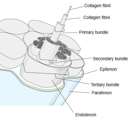
Glossary of terms
| Term | Explanation | |
|---|---|---|
| 1 | Collagen fibre | the largest component of the tendon |
| 2 | Collagen fibril | the unit of the collagen fibre: many collagen molecules, each a triple helix of three polypeptide chains, lying side by side |
| 3 | Tendon bundle | collagen fibres wrapped by the endotenon: primary, secondary and tertiary bundles |
| 4 | Endotenon | loose connective tissue around each bundle, carrying blood vessels, nerves and lymphatics |
| 5 | Epitenon | connective tissue around the whole tendon, carrying blood vessels, nerves and lymphatics |
| 6 | Paratenon | the outermost connective-tissue layer around the tendon, rich in blood vessels; it lets the tendon glide |
| 7 | Peritendineum | an older term for the connective-tissue sheaths of the tendon (internal and external peritendineum); sources differ on how it matches the endotenon and epitenon |
| 8 | Tenocyte | the cell of the tendon; tenocytes and tenoblasts make up most of the cells, the rest are chondrocyte-like cells |
Other points
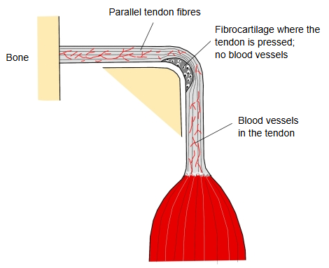
Where a tendon bends around a bone and is pressed against it, fibrocartilage forms in the tendon and there are no blood vessels.
At the attachment to bone, some of the collagen fibres of the tendon pass through the periosteum into the bone (perforating fibres) and anchor the tendon firmly.
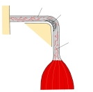 Tendon bending around a bone
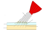 Tendon attachment to bone
Tendons seen under the skin
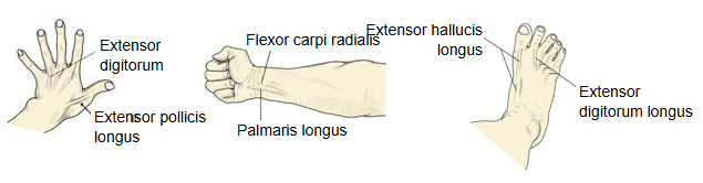
Muscles shown: extensor digitorum, extensor pollicis longus, flexor carpi radialis, palmaris longus, extensor hallucis longus, extensor digitorum longus. The calcaneal tendon can also be seen and felt under the skin.
Reference websites
[Wikipedia]
· Tendon, Wikipedia.
Related terms
- Calcaneal tendon
- Central tendon of diaphragm
- Palmar aponeurosis
- Epicranial aponeurosis
- Synovial membrane
- Extensor digitorum
- Palmaris longus
References and sources
- Further reading: Tendon, Wikipedia.
- Federative International Programme for Anatomical Terminology (FIPAT). Terminologia Anatomica, 2nd ed. (TA2), 2019.