Median palatine suture (Latin: sutura palatina mediana)
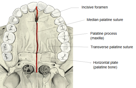
The median palatine suture is the joint between the lower, medial edges of the left and right maxillae (palatine processes) and the left and right palatine bones (horizontal plates).
Maxilla and palatine bone: both are paired bones of the facial skeleton (viscerocranium)
In other words, it is the suture line running from front to back along the roof of the oral cavity; near its front end is the incisive fossa, where the incisive foramen, the outlet of the incisive canal, opens.
Roof of the oral cavity = palateIncisive canal: the passage for nerves and vessels between the nasal cavity and the mouth
On the opposite side of the median palatine suture, the nasal side, it forms a raised line, the nasal crest, which ends in front by projecting forwards as the anterior nasal spine.
Nasal side: the surface facing the nasal cavityNasal crest and anterior nasal spine: the cartilaginous part of the nasal septum is attached here
· The palatal shelves of the two sides fuse in the midline at about weeks 9 to 12 of fetal life, starting at the front.
· The suture is not ossified and stays flexible through childhood and the early teens, which is why orthodontic palatal expansion works best in children. It ossifies only slowly, starting at the back, from the end of growth into adulthood; the timing varies greatly, and in some adults the suture is still partly open.
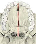 Median palatine suture
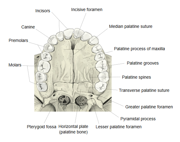
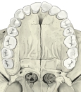 Hard palate and teeth (inferior view)
Reference websites
[Wikipedia]
· Hard palate, Wikipedia.
· Palatal expansion, Wikipedia.
[Websites with illustrations]
· Bony palate from below (Sobotta 1909, fig. 100) (Wikimedia Commons)

Related terms
- Incisive fossa
- Incisive foramen
- Incisive bone
- Incisive papilla
- Incisive crest
- Transverse palatine suture
- Palatine process
- Horizontal plate
- Bony palate
- Hard palate
- Incisive canal
- Maxilla
References and sources
- Further reading: Hard palate, Wikipedia.
- Federative International Programme for Anatomical Terminology (FIPAT). Terminologia Anatomica, 2nd ed. (TA2), 2019.