Incisive papilla (Latin: papilla incisiva)
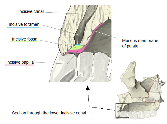
It lies in the midline at the front of the hard palate, over the incisive fossa, where the incisive canals open onto the palate, and it marks the position of the fetal nasopalatine canal. The transverse palatine folds spread out to each side just behind it. It is a pad of dense connective tissue covered by the firm, often keratinised mucosa of the hard palate. It is a landmark in dentistry, for example for anaesthetising the nasopalatine nerve and for positioning the front teeth of dentures.
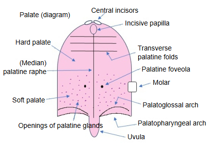
Below is a simple outline of the features of the hard palate and the soft palate.
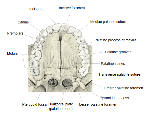
- Hard palate : the front part, with a base of bone (the palatine processes of the maxillae and the horizontal plates of the palatine bones) covered by mucosa. Seen on its surface: the palatine raphe, the transverse palatine folds, the incisive papilla and others.
- Soft palate : the back part, with a base of muscle covered by mucosa; it hangs down behind as the uvula. Its muscles: palatopharyngeus, musculus uvulae, palatoglossus, levator veli palatini, tensor veli palatini.
 Lower part of the incisive canal (section)
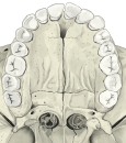 Bony palate (inferior view)
Reference websites
[Wikipedia]
· Incisive papilla, Wikipedia.
· Hard palate, Wikipedia.
[Websites with illustrations]
· The palate (labelled drawing) (Wikimedia Commons)

· Features of the hard palate (figure) (Reddit)

Related terms
- Incisive fossa
- Incisive foramen
- Incisive bone
- Incisive crest
- Median palatine suture
- Hard palate
- Palatine raphe
- Transverse palatine folds
- Incisive canal
- Incisor
- Palate
- Nasopalatine nerve
References and sources
- Further reading: Incisive papilla, Wikipedia.
- Further reading: Hard palate, Wikipedia.
- Federative International Programme for Anatomical Terminology (FIPAT). Terminologia Anatomica, 2nd ed. (TA2), 2019.