Palate (Latin: palatum)
- Overview
- Nerves and vessels
- Terms related to the palate
- Reference websites
- (Wikipedia, websites with illustrations)
- Where it is found
- Related terms
Overview
nology
- Respiratory system : nose, pharynx, larynx, trachea, bronchi, lungs
- Digestive system
- Oral cavity : lips, oral cavity, palate, floor of mouth, cheeks (the oral cavity as a part of the digestive system)
- Pharynx : pharynx
- Oesophagus : then the stomach, the small intestine and the large intestine
- Other systems : urinary system, reproductive system, peritoneum, endocrine glands
cavity
- Lips : the front wall of the mouth: the upper lip and lower lip, joined at each corner (the labial commissure).
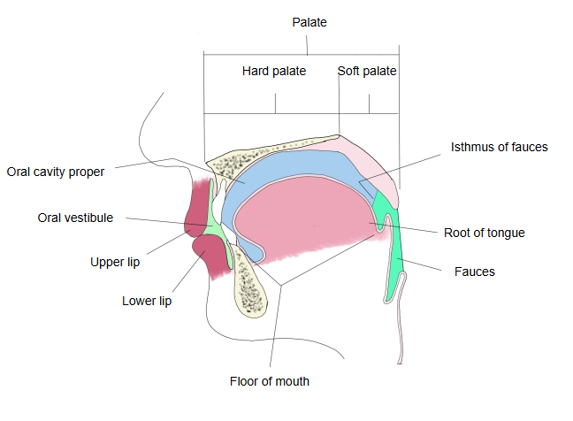
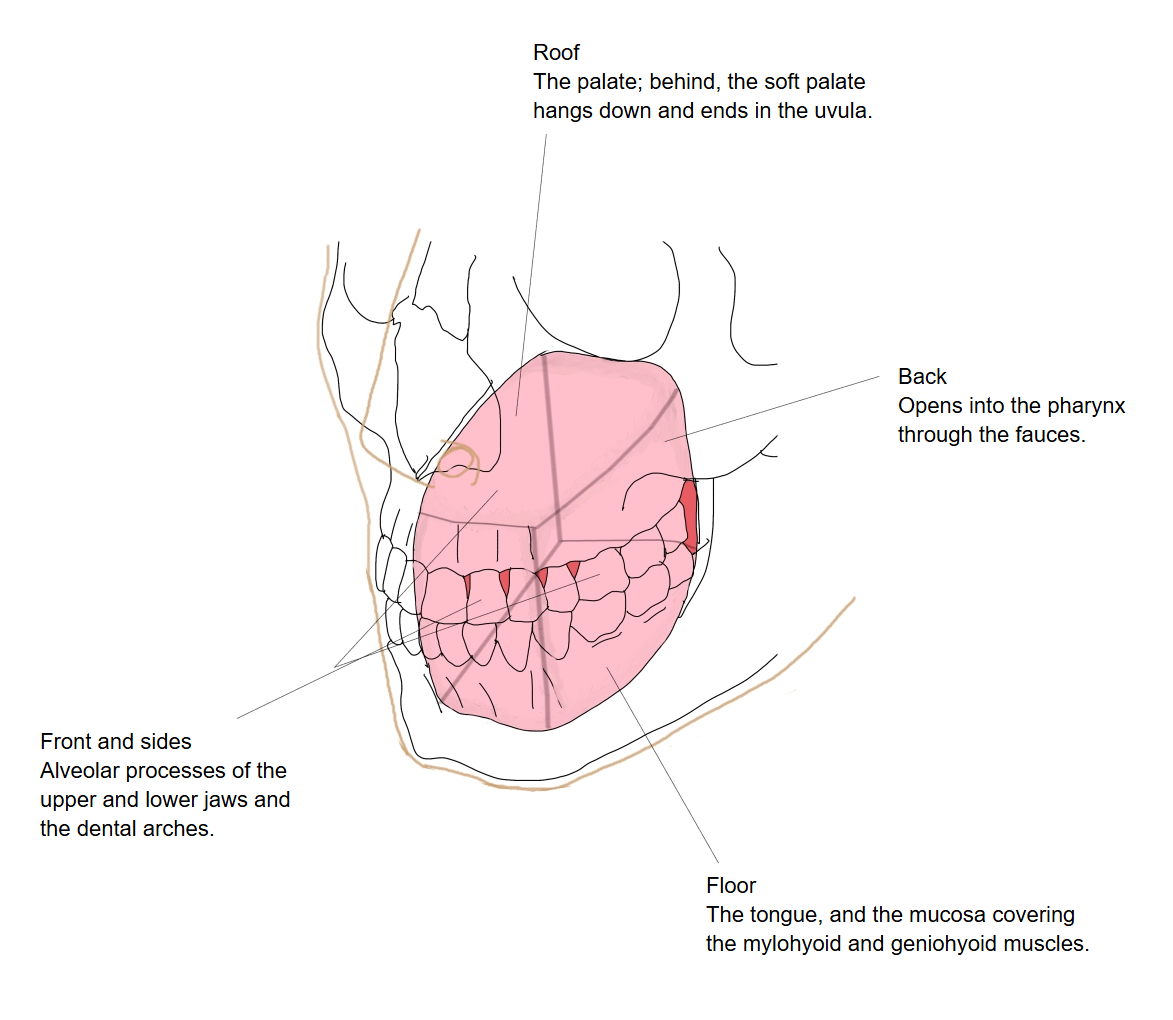
- Oral cavity : the space inside the lips and cheeks, in two parts: the oral vestibule in front (narrow) and the oral cavity proper behind.
- Palate : the upper wall (roof) of the mouth, in two parts: the hard palate in front (the larger part) and the soft palate behind.
- Floor of mouth : the bottom of the mouth, covered by soft mucous membrane with much loose connective tissue beneath.
- Cheeks : the side walls of the mouth; they form the oral vestibule with the lips and are made of skin, muscle (buccinator) and mucous membrane.
of the
palate
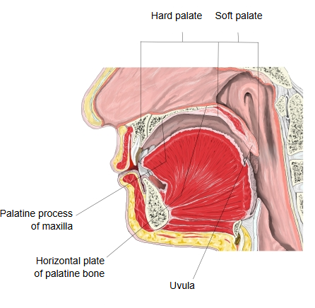
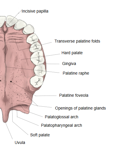
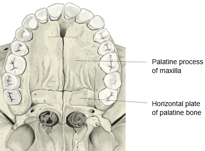
- Hard palate : the front part, with bone as its base (palatine processes and horizontal plates), covered by mucous membrane. Seen on its surface: the palatine raphe, the transverse palatine folds, the incisive papilla and others.
- Soft palate : the back part, with muscle as its base, covered by mucous membrane; the part hanging down from it is the uvula. Its muscles: palatopharyngeus, musculus uvulae, palatoglossus, levator veli palatini and tensor veli palatini.
The palate separates the oral cavity below from the nasal cavity and the nasal part of the pharynx above; it forms the roof of the oral cavity proper and is bounded in front and at the sides by the upper gums and teeth. Its mucous membrane contains many small palatine glands, most of them toward the back.
The soft palate is a sheet of muscle and fibrous tissue (the palatine aponeurosis) covered by mucous membrane; two small pits near the midline, the palatine foveolae, lie close to where it joins the hard palate. Two folds run down from each side of it: the palatoglossal arch in front and the palatopharyngeal arch behind.
The palate works with the tongue in chewing, swallowing and speech; in swallowing the soft palate rises and closes off the nasal part of the pharynx. If its halves fail to fuse during development, the result is a cleft palate.
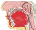 Mouth and pharynx (median section)
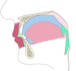 Oral cavity (median section, diagram)
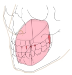 Oral cavity proper
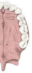 Mucosa of the palate
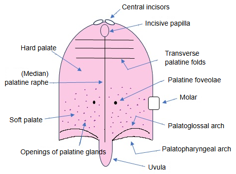 Mucosa of the palate (diagram)
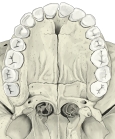 Bony palate (inferior view)
Nerves and vessels
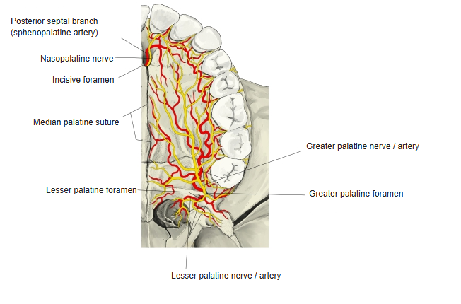
Nerves: the greater palatine nerve ⇒ hard palate (gums, mucous membrane, glands)
The nasopalatine nerve ⇒ hard palate (mucous membrane behind the incisor teeth)
The lesser palatine nerves ⇒ soft palate (sensory: the palatine tonsil and the lower part of the velum palatinum; some older texts also give them motor fibres to levator veli palatini and musculus uvulae; sources differ)
Arteries: the greater palatine artery ⇒ hard palate
The lesser palatine arteries ⇒ soft palate
Veins: companion veins of these arteries, the greater palatine vein and the lesser palatine veins.
The sensory nerves are branches of the maxillary nerve that pass through the pterygopalatine ganglion. The muscles of the soft palate are supplied through the pharyngeal plexus (vagus nerve), except tensor veli palatini, which is supplied by the mandibular nerve.
The greater and lesser palatine arteries both come from the descending palatine artery, a branch of the maxillary artery. The soft palate also receives the ascending palatine branch of the facial artery.
Terms related to the palate
- Bony palate
- Hard palate
- Horizontal plate
- Incisive canal
- Incisive fossa
- Incisive papilla
- Palatine process
- Palatine raphe
- Palatoglossal arch
- Palatopharyngeal arch
- Soft palate
- Transverse palatine folds
- Uvula
Reference websites
[Wikipedia]
· Palate, Wikipedia.
· Hard palate, Wikipedia.
· Soft palate, Wikipedia.
[Websites with illustrations]
· The palate (TeachMeAnatomy)
· Palate: anatomy, innervation, blood supply and function (Kenhub)
Where it is found
Related terms
- Hard palate
- Soft palate
- Palatine process
- Palatine bone
- Palatoglossal arch
- Palatopharyngeal arch
- Palatine glands
References and sources
- Further reading: Palate, Wikipedia.
- Further reading: Hard palate, Wikipedia.
- Further reading: Soft palate, Wikipedia.
- Federative International Programme for Anatomical Terminology (FIPAT). Terminologia Anatomica, 2nd ed. (TA2), 2019.