Outer hair cell
of
Corti
- Hair cells
- Inner hair cells : in a single row on the inner side of the inner pillar cells, with stereocilia on their free surface; about 3,500.
- Outer hair cells : in several rows on the outer side of the outer pillar cells; about 12,000, three to four times as many as the inner hair cells.
- Supporting cells
- Inner pillar cells : outside the inner hair cells; with the outer pillar cells they form the tunnel of Corti.
- Outer pillar cells : inside the outer hair cells; with the inner pillar cells they form the tunnel of Corti.
- Inner phalangeal cells : on the inner side of the inner pillar cells; they support the inner hair cells.
- Outer phalangeal cells : = cells of Deiters. Outside the outer pillar cells; they support the outer hair cells, in rows matching them. Related terms: space of Nuel, phalangeal process, reticular membrane, Retzius fibre.
- Cells of Hensen : several rows of cells outside the outer phalangeal cells, forming the outer slope of the organ of Corti.
Features
· The cells are arranged alternately (staggered) both across and along the rows.
· Their shape resembles that of the inner hair cells, but there are three to four times as many of them.
· The number and arrangement of the stereocilia on the outer hair cells differ from turn to turn of the cochlea.
· The cells stand almost at right angles to the surface of the reticular membrane, and make an acute angle, open inwards, with the basilar membrane.
· The top of each cell fits into its own opening in the reticular membrane and is held there.
· The basic shape is a cylinder, but the lower half (which holds a large round nucleus) is slightly swollen, close to a cone.
· The lower end does not reach the basilar membrane: it sits in the cup of an outer phalangeal cell (cell of Deiters), at its neck.
· The number of stereocilia differs from row to row; there are about a hundred on each cell, standing in a V- or W-shaped pattern, and the tallest are embedded in the tectorial membrane.
· The number of rows differs between the turns of the cochlear duct:
First (basal) turn: usually three rows, with a cell missing here and there.
Second (middle) turn: usually four rows; as in the first turn, a cell is missing here and there.
Third (apical) turn: an incomplete fifth row appears, made of scattered single cells.
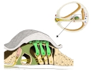 Spiral organ (section)
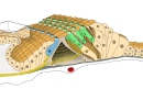 Spiral organ (3D view)
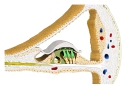 Cochlear duct (section)
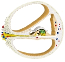 Section of a cochlear turn
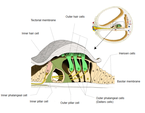
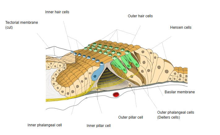
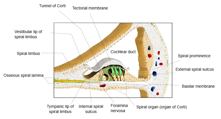
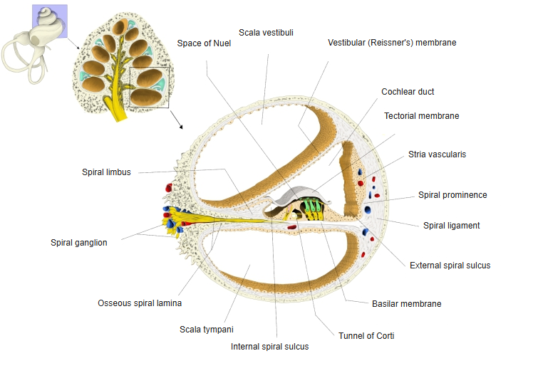
Function and further details
When stimulated, the outer hair cell shortens and lengthens in step with the sound (electromotility, driven by the motor protein prestin). This "cochlear amplifier" makes the ear more sensitive to quiet sounds and sharpens the tuning of each place on the basilar membrane. The cells receive mainly efferent nerve fibres from the brainstem, which adjust the amplification; only a few afferent fibres of the cochlear nerve carry signals from them, and the nerve endings are densest at the base of the cell. Older descriptions also mention a dark, rounded body in the upper half of the cell (Hensen's body, or spiral body), and note that the number of rows varies between individuals; the inner hair cells, normally in a single row, sometimes show a second row here and there.
Reference websites
[Wikipedia]
· Hair cell, Wikipedia.
· Cochlear amplifier, Wikipedia.
[Websites with illustrations]
· Organ of Corti (diagram) (Wikimedia Commons)

Related terms
- Organ of Corti
- Outer phalangeal cell
- Reticular membrane
- Tunnel of Corti
- Basilar membrane
- Cochlear duct
- Cochlear nerve
References and sources
- Further reading: Hair cell, Wikipedia.
- Further reading: Cochlear amplifier, Wikipedia.
- Federative International Programme for Anatomical Terminology (FIPAT). Terminologia Anatomica, 2nd ed. (TA2), 2019.