Ciliary muscle (Latin: musculus ciliaris)
ocular
muscles
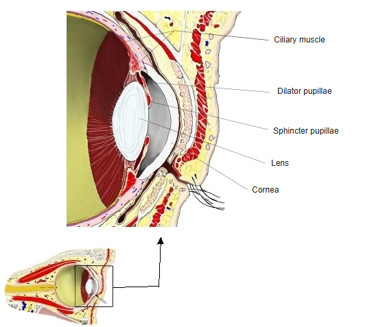
- Ciliary muscle : surrounds the lens and adjusts its thickness
- Sphincter pupillae : runs in a ring in the iris and narrows the pupil; one of the iris muscles
- Dilator pupillae : runs radially at the outer part of the iris and widens the pupil; one of the iris muscles
· The ciliary muscle is one of the intra-ocular muscles inside the eyeball, lying in the ciliary body.
· It is supplied by parasympathetic fibres of the oculomotor nerve (CN III): preganglionic fibres from the accessory oculomotor (Edinger–Westphal) nucleus relay in the ciliary ganglion, and the postganglionic fibres reach the muscle in the short ciliary nerves. The same pathway supplies the sphincter pupillae.
nerve
(CN III)
- From the oculomotor nucleus : motor fibres; the nerve leaves the midbrain on the medial side of the cerebral peduncle and enters the orbit through the superior orbital fissure
- Superior branch : superior rectus, levator palpebrae superioris
- Inferior branch : medial rectus, inferior rectus, inferior oblique
- From the accessory oculomotor (Edinger–Westphal) nucleus : parasympathetic fibres, which leave with the inferior branch
- Ciliary ganglion : relay; then the short ciliary nerves to the sphincter pupillae and the ciliary muscle
muscle
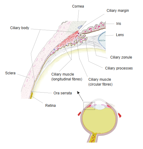
- Circular fibres : = Müller's muscle: the innermost ring, in the front and inner part of the muscle near the root of the iris; its size varies widely between individuals
- Longitudinal (meridional) fibres : = Brücke's muscle: the outer fibres, running from front to back just inside the sclera from the region of the junction of the cornea and sclera into the ciliary body
- Radial (oblique) fibres : between the circular and longitudinal fibres, fanning out from the scleral spur near the corneoscleral junction
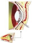 Intra-ocular muscles
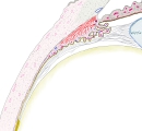 Ciliary body (section)
Reference websites
[Wikipedia]
· Ciliary muscle, Wikipedia.
[Websites with illustrations]
· Ciliary muscle (Kenhub)
· Section through the front of the eyeball with the radial and circular fibres of the ciliary muscle (Gray's Anatomy) (Wikimedia Commons)

· The three fibre groups of the ciliary muscle (longitudinal, radial and circular) (American Academy of Ophthalmology)
Development and nerve supply
The ciliary muscle develops from mesenchyme of the developing choroid, which is regarded as derived from the cranial neural crest. Its nerve supply is parasympathetic: preganglionic fibres from the accessory oculomotor (Edinger–Westphal) nucleus in the midbrain travel in the oculomotor nerve and its inferior branch to the ciliary ganglion; postganglionic fibres reach the muscle in the short ciliary nerves, which run with branches of the nasociliary nerve. The transmitter, acetylcholine, acts on muscarinic (M3) receptors and makes the muscle contract. Parasympathetic tone is high when the eye focuses on near objects, as in reading.
Function
Accommodation: when the ciliary muscle contracts, its ring narrows and moves forwards and inwards towards the lens. The ciliary zonule slackens and the elastic lens becomes rounder and more strongly refracting, for near vision. When the muscle relaxes, the zonule tightens and the lens flattens, for distant vision. This is the classic account (Helmholtz); sources differ on some details, and other theories have been proposed. Long periods of close work, such as reading fine print, tire the eyes mainly through fatigue of the ciliary muscle. Outflow of aqueous humour: the longitudinal fibres pull on the scleral spur and the trabecular meshwork; their contraction opens the spaces of the meshwork and helps aqueous humour drain into the scleral venous sinus (canal of Schlemm).
Related terms
- Intraocular muscles
- Ciliary body
- Meridional fibres of ciliary muscle
- Radial fibers of ciliary muscle
- Circular fibres of ciliary muscle
- Ciliary zonule
- Muscles of iris
- Lens
References and sources
- Further reading: Ciliary muscle, Wikipedia.
- Federative International Programme for Anatomical Terminology (FIPAT). Terminologia Anatomica, 2nd ed. (TA2), 2019.