Flexor retinaculum of foot (Latin: retinaculum musculorum flexorum pedis)
The flexor retinaculum of the foot is the fibrous band stretched between the medial malleolus of the tibia and the calcaneus. It lies at the ankle; the flexor retinaculum of the wrist is a different structure. The following pass beneath it.
| Tendons | 1 | Tendon of tibialis posterior |
| 2 | Tendon of flexor hallucis longus | |
| 3 | Tendon of flexor digitorum longus | |
| Vessel | 1 | Posterior tibial artery |
| Nerves | 1 | Lateral plantar nerve |
| 2 | Medial plantar nerve |
Some sources list the tibial nerve here instead, since it divides into the two plantar nerves at about this level.
Its upper border is continuous with the deep fascia of the leg, and its lower border with the plantar aponeurosis and the fibres of origin of abductor hallucis. Together with the grooves in the bones beneath, it forms the tarsal tunnel, through which these structures reach the sole.
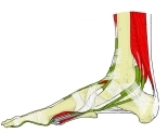 Right foot (medial view)
 Arteries and nerves of the right foot (medial view)
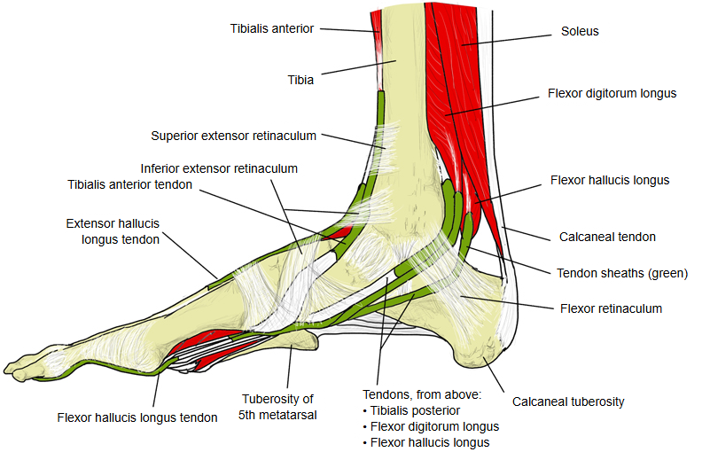
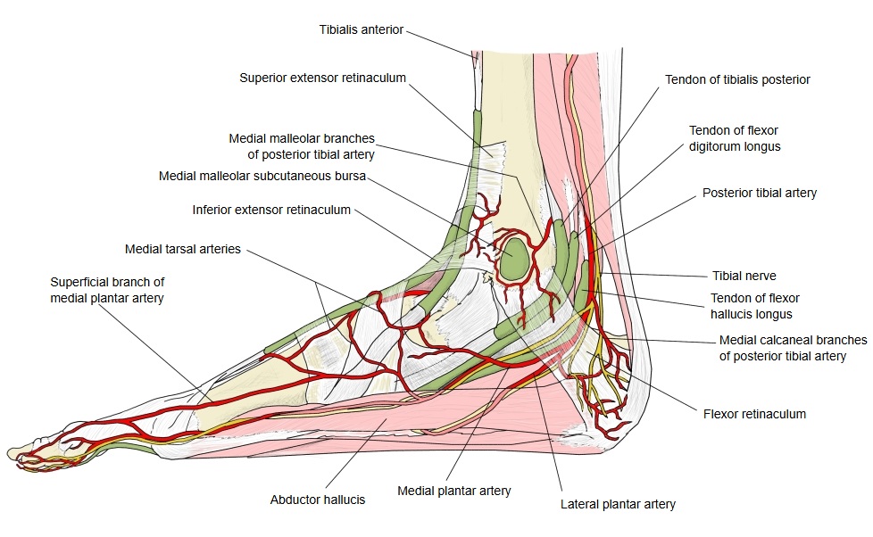
Reference websites
[Wikipedia]
· Flexor retinaculum of foot, Wikipedia.
· Tarsal tunnel, Wikipedia.
[Websites with illustrations]
· Retinacula and tendon sheaths of the ankle (drawing) (Anatomy Facts)
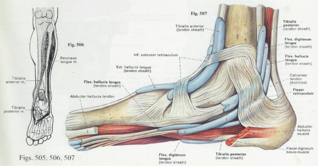
Related terms
- Medial malleolus
- Calcaneus
- Tibialis posterior
- Flexor digitorum longus
- Flexor hallucis longus
- Posterior tibial artery
- Tibial nerve
References and sources
- Further reading: Flexor retinaculum of foot, Wikipedia.
- Federative International Programme for Anatomical Terminology (FIPAT). Terminologia Anatomica, 2nd ed. (TA2), 2019.