Latin: patella · also called the kneecap
Overview
- A bone of the free lower limb at the front of the knee.
- The largest sesamoid bone of the body: it develops in the tendon of the quadriceps femoris.
- A flat, triangular bone shaped like a chestnut: the broad base faces upwards and the pointed apex downwards.
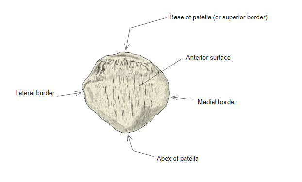
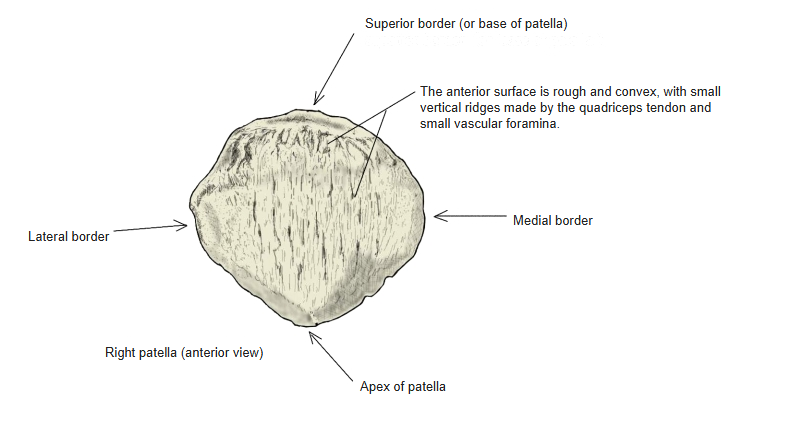
- It has 2 surfaces (anterior and posterior) and 3 borders (superior, medial and lateral). The superior border is also called the base of patella.
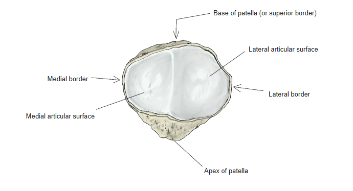
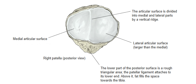
- The posterior surface carries the articular surface, divided into medial and lateral facets; this articular surface glides on the patellar surface of the femur.
Bones of the lower limb 
Knee region (anterior)
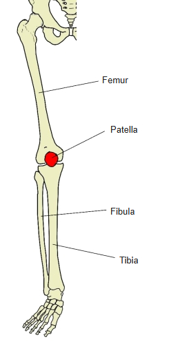
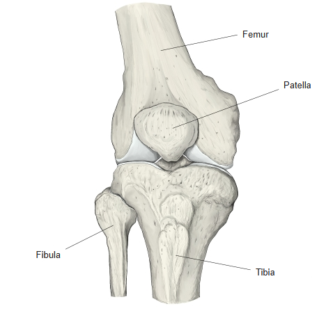
- Bones of the lower limb (one side)
- Pelvic girdle
- Hip bone ← ilium, ischium and pubis fused
- Free lower limb
- Thigh and knee
- Femur
- Patella
- Leg
- Tibia ← big-toe side
- Fibula ← little-toe side
- Foot
- Tarsal bones (7)
- Metatarsal bones (5)
- Phalanges (14)
- Thigh and knee
- Pelvic girdle
Illustrations
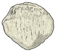
Right patella (anterior) 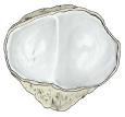
Right patella (posterior) 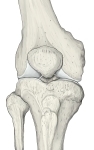
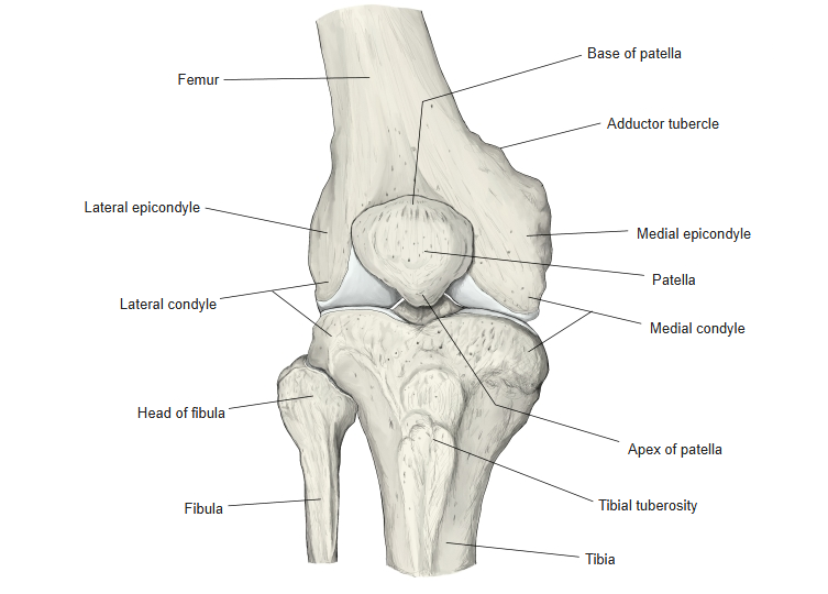
Right knee (anterior) 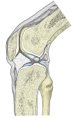
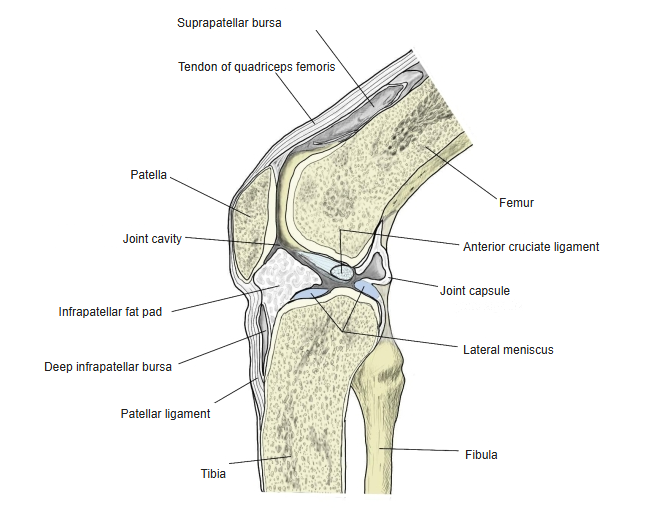
Right knee joint (sagittal section)
Articulations
| # | Bone | Joint |
|---|---|---|
| 1 | Femur | Knee joint |
Patellofemoral surfaces
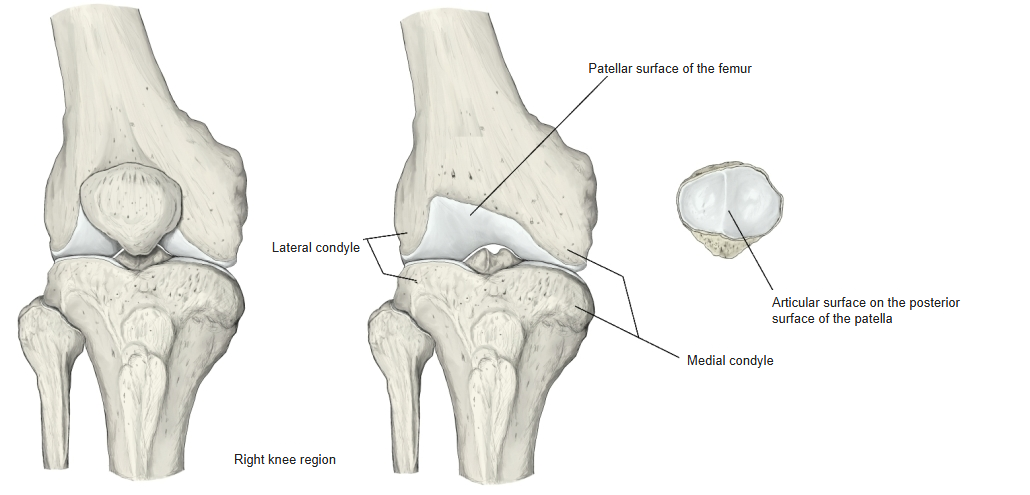
Knee joint: articular surface on the posterior surface of the patella ↔ patellar surface of the femur. Some sources name this part the patellofemoral joint.
Ligaments
The following ligaments attach to the patella. The list is intended to be comprehensive but may omit minor ligaments.
| # | Ligament | Connects |
|---|---|---|
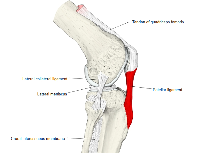
| 1 | Patellar ligament | Lower part (apex) of the patella – tibial tuberosity |
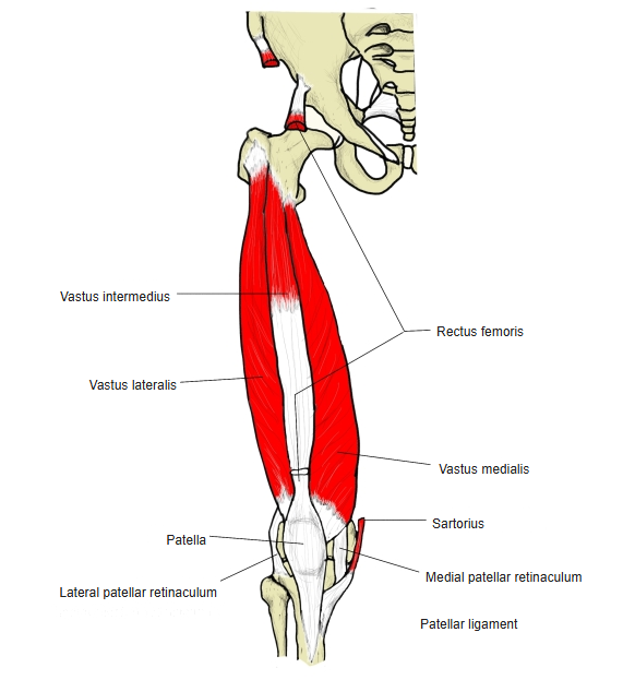
The patella is stabilised by the horizontal fibres of the vastus medialis and by the prominence of the lateral condyle of the femur, which resists lateral dislocation during flexion; the patellar retinacula also stabilise it during movement.
Patellar ligament
Muscle attachments
- Origin of: none
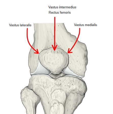
- Insertion of: Rectus femoris, Vastus intermedius, Vastus medialis, Vastus lateralis
All four heads of the quadriceps femoris insert on the patella through the quadriceps tendon, and continue to the tibial tuberosity through the patellar ligament.
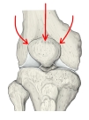
Quadriceps insertion 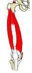
Quadriceps femoris
Surfaces and borders
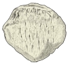
- Anterior surface: rough and convex, with small vertical ridges made by the quadriceps tendon and small vascular foramina.
- Posterior surface: the smooth articular surface, divided by a vertical ridge into a medial and a larger lateral part; below it a rough triangular area where the patellar ligament attaches, with the infrapatellar fat pad lying between the ligament and the tibia.
- Borders: superior (base of patella), medial and lateral; the lower point is the apex of patella.
Anterior surface 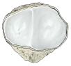
Posterior surface
Development and variations
- Ossification begins at about 3 years and is complete at about 20 years.
- It may be absent, or divided into an upper and a lower part joined by a ligament.
- Emarginations (a “missing piece”, patella emarginata) are common at the lateral part of the upper edge.
- A bipartite patella results from ossification of a second cartilaginous layer at the site of an emargination (formerly explained as failure of ossification centres to fuse). Partite patellas are much more common in males than in females; tripartite and multipartite forms also occur.
Blood supply
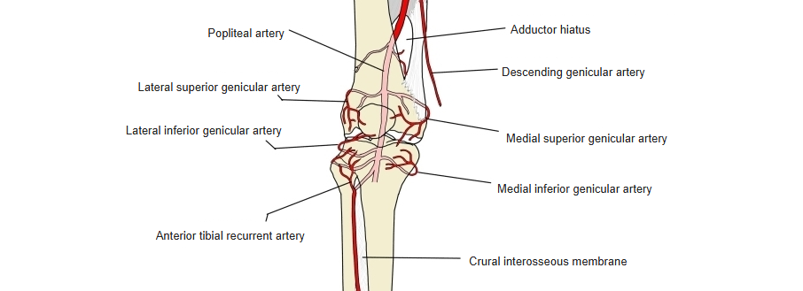
- From the arterial network around the knee (genicular anastomosis); probably mainly the lateral and medial superior genicular arteries (not certain).
- Other contributions described include the recurrent branch of the anterior tibial artery and the descending branch of the lateral circumflex femoral artery.
Arteries around the knee
References and sources
- Wheeless' Textbook of Orthopaedics: Blood supply to the knee.
- Further reading: Patella, Wikipedia.