Latin: os ethmoidale
Overview
- An unpaired, very light bone at the front of the cranial base, lying between the two orbits and forming the roof and much of the walls of the nasal cavity.
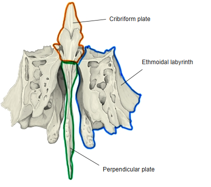
- It has three parts: the horizontal cribriform plate, the midline perpendicular plate (with the crista galli projecting upwards) and the paired ethmoidal labyrinths filled with ethmoidal air cells.
- Ossification begins in the cartilage of the nasal capsule before birth; the three parts fuse at about six years of age.
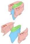
Parts of the ethmoid
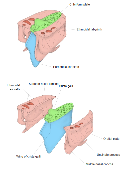
- Bones of the skull
- Neurocranium (cranial bones)
- Frontal bone
- Parietal bone (2)
- Occipital bone
- Temporal bone (2)
- Sphenoid bone
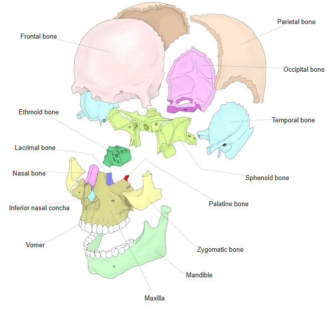
- Ethmoid bone
- Viscerocranium (facial bones)
- Nasal bone (2)
- Vomer
- Lacrimal bone (2)
- Inferior nasal concha (2)
- Maxilla (2)
- Zygomatic bone (2)
- Palatine bone (2)
- Mandible
- Hyoid bone
- Neurocranium (cranial bones)
Illustrations
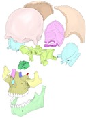
Bones of the skull (separated) 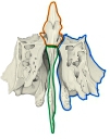
Ethmoid (anterior) 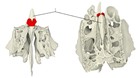
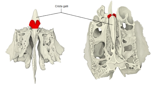
Crista galli
Articulations
| # | Bone | Joint |
|---|---|---|
| 1 | Frontal bone | Ethmoidal notch and nasal part; the wing of crista galli meets the nasal part |
| 2 | Sphenoid bone | Front of the body of the sphenoid |
| 3 | Nasal bones | Perpendicular plate |
| 4 | Maxillae | Frontal process and orbital surface |
| 5 | Lacrimal bones | Orbital plate |
| 6 | Palatine bones | Labyrinth |
| 7 | Vomer | Perpendicular plate |
| 8 | Inferior nasal conchae | Ethmoidal process |
| 9 | Septal cartilage of the nose | Perpendicular plate |
Sagittal section of the nasal cavity 
Internal cranial base
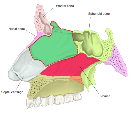
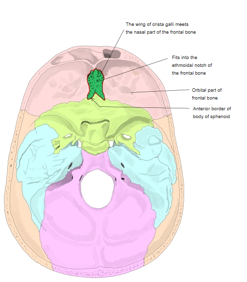
The ethmoid is outlined in red in the sagittal section and shown in green in the cranial base.
Muscle attachments
- Origin of: none
- Insertion of: none
No muscles attach to the ethmoid.
Main features
- Cribriform plate – a horizontal sieve-like plate in the anterior cranial fossa; its holes carry the olfactory nerve fibres. The crista galli rises from its midline, with the small wings of the crista galli at its front, and the foramen cecum lies in front of it.
- Perpendicular plate – a thin midline plate forming the upper part of the nasal septum; it meets the vomer and the septal cartilage below.
- Ethmoidal labyrinth – a box of thin-walled ethmoidal air cells (anterior, middle and posterior), together called the ethmoidal sinus. Its lateral wall is the smooth orbital plate, part of the medial wall of the orbit; the anterior and posterior ethmoidal foramina open at its upper edge.
- Nasal conchae – the superior and middle nasal conchae hang from the medial side of the labyrinth, roofing the superior and middle nasal meatuses. A small supreme nasal concha is sometimes present above the superior concha.
- Ethmoidal bulla, uncinate process and ethmoidal infundibulum – features in the middle meatus where the anterior air cells and frontal and maxillary sinuses drain.
References and sources
- Further reading: Ethmoid bone, Wikipedia.