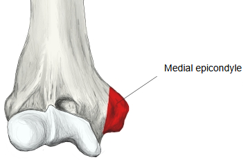
Medial epicondyle (Latin: epicondylus medialis humeri)
Also called the medial epicondyle of humerus
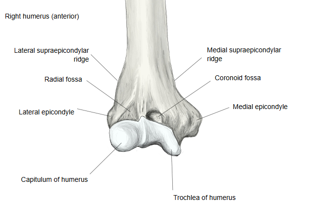
- It is mainly the place where the flexor muscles of the forearm arise (the common flexor origin): pronator teres (humeral head), flexor carpi radialis, palmaris longus, flexor carpi ulnaris and flexor digitorum superficialis.
- The femur also has a projection with the same name, the medial epicondyle of the femur.
- On the opposite, lateral side is the lateral epicondyle, which projects less than the medial epicondyle.
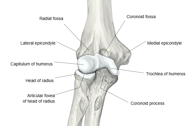
Being larger and more prominent than the lateral epicondyle, it is easy to feel at the inner side of the elbow. The ulnar collateral ligament of the elbow is attached to it. The ulnar nerve runs in the groove for the ulnar nerve on its back, close to the skin; a knock here gives the tingling of the "funny bone". Above it, the medial supracondylar ridge runs up the shaft.
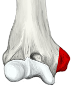 Medial epicondyle (red)
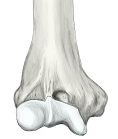 Lower end (anterior)
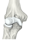 Right elbow (anterior)
Reference websites
[Wikipedia]
· Medial epicondyle of the humerus, Wikipedia.
[Websites with illustrations]
· Left humerus, anterior view (Gray's Anatomy, fig. 207) (Wikimedia Commons)

· Left humerus, posterior view (Gray's Anatomy, fig. 208) (Wikimedia Commons)

· The humerus: proximal, shaft and distal parts (TeachMeAnatomy)
Where it is found
Related terms
- Lateral epicondyle
- Medial supracondylar ridge
- Groove for ulnar nerve
- Trochlea of humerus
- Distal epiphysis of humerus
- Medial epicondyle of femur
References and sources
- Further reading: Medial epicondyle of the humerus, Wikipedia.
- Federative International Programme for Anatomical Terminology (FIPAT). Terminologia Anatomica, 2nd ed. (TA2), 2019.