Forearm flexor muscles
Also called the flexor muscles of forearm or the anterior compartment muscles of forearm
The list below follows sources that divide the forearm flexors into four layers. Many English texts use three layers (superficial, intermediate and deep), counting pronator quadratus with the deep layer.
flexor
muscles
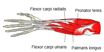
- Layer 1 : Pronator teres, flexor carpi radialis, palmaris longus, flexor carpi ulnaris
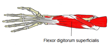
- Layer 2 : Flexor digitorum superficialis
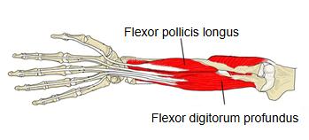
- Layer 3 : Flexor pollicis longus, flexor digitorum profundus
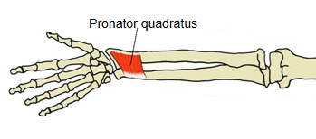
- Layer 4 : Pronator quadratus
The table below describes these muscles in a little more detail, but the origins, insertions and actions are much simplified. See the page of each muscle for details.
| Layer | No | Name | Origin | Insertion | Action |
|---|---|---|---|---|---|
| Layer 1 | 1 | Pronator teres | Medial epicondyle of the humerus; coronoid process of the ulna | Pronator tuberosity of the radius | Pronates the forearm; helps flex the elbow |
| 2 | Flexor carpi radialis | Medial epicondyle of the humerus | Bases of the 2nd and 3rd metacarpals | Flexes and abducts the wrist; helps pronate the forearm | |
| 3 | Palmaris longus | Medial epicondyle of the humerus | Palmar aponeurosis | Flexes the wrist; often absent on one or both sides | |
| 4 | Flexor carpi ulnaris | Medial epicondyle of the humerus; posterior border of the ulna | Pisiform, hook of hamate and base of the 5th metacarpal | Flexes and adducts the wrist | |
| Layer 2 | 5 | Flexor digitorum superficialis | Medial epicondyle of the humerus, coronoid process of the ulna, front of the radius | Bases of the middle phalanges of the 2nd–5th fingers | Flexes the proximal interphalangeal joints of the index to little fingers, then the knuckles and wrist |
| Layer 3 | 6 | Flexor pollicis longus | Front of the radius and the interosseous membrane | Distal phalanx of the thumb | Flexes the thumb, especially its interphalangeal joint |
| 7 | Flexor digitorum profundus | Upper part of the front and medial surface of the ulna; interosseous membrane | Bases of the distal phalanges of the 2nd–5th fingers | Flexes the distal interphalangeal joints (the only muscle that does); helps flex the other finger joints and the wrist | |
| Layer 4 | 8 | Pronator quadratus | Lower quarter of the front of the ulna | Lower quarter of the front of the radius | The main pronator of the forearm |
Most of them arise by a common flexor origin from the medial epicondyle of the humerus; the deeper ones arise from the front of the radius, the ulna and the interosseous membrane. All are supplied by the median nerve, except flexor carpi ulnaris and the medial (ulnar) half of flexor digitorum profundus, which are supplied by the ulnar nerve. Their long tendons cross the front of the wrist; the finger and thumb flexor tendons pass through the carpal tunnel under the flexor retinaculum. On the back of the forearm lie the forearm extensor muscles.
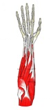 Layer 1 (superficial layer)
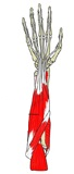 Layer 2 (intermediate layer)
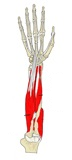 Layer 3 (deep layer)
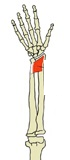 Layer 4 (deepest layer)
Reference websites
[Wikipedia]
· Anterior compartment of the forearm, Wikipedia.
[Websites with illustrations]
· Anterior forearm flexor muscles (TeachMeAnatomy)
· Cross-section through the middle of the forearm (Gray's Anatomy, fig. 417) (Wikimedia Commons)

Related terms
- Forearm extensor muscles
- Interosseous membrane of forearm
- Medial epicondyle
- Carpal tunnel
- Flexor retinaculum (hand)
- Pronator teres
- Flexor digitorum superficialis
- Flexor digitorum profundus
- Flexor carpi ulnaris
References and sources
- Further reading: Anterior compartment of the forearm, Wikipedia.
- Federative International Programme for Anatomical Terminology (FIPAT). Terminologia Anatomica, 2nd ed. (TA2), 2019.