Body of radius (Latin: corpus radii)
Also called the shaft of radius
The body is a three-sided prism with three surfaces (anterior, posterior and lateral) and three borders (anterior, posterior and the medial border, which is the interosseous border). It is narrower above and wider below, and is slightly bowed so that it is convex laterally. Its lower part widens towards the ulna, and the gap between the two bones (the interosseous space) is wide in the middle and lower part of the forearm.
- Anterior surface : between the anterior and interosseous borders.
- Posterior surface : between the posterior and interosseous borders.
- Lateral surface : between the anterior and posterior borders.
- Anterior border : between the anterior and lateral surfaces.
- Posterior border : between the posterior and lateral surfaces.
- Medial border : the sharp border facing the ulna. = interosseous border
Related features of the body: radial tuberosity (near the upper end, on the medial side) · pronator tuberosity (about the middle of the lateral surface)
| Muscles attached to the body of the radius | |
|---|---|
| Origin | Flexor digitorum superficialis (radial head) · Flexor pollicis longus · Abductor pollicis longus · Extensor pollicis brevis |
| Insertion | Biceps brachii (radial tuberosity) · Supinator · Pronator teres · Pronator quadratus |
Not on the body: brachioradialis is inserted on the lower end of the radius (distal epiphysis), just above the styloid process.
- Proximal epiphysis : the disc-shaped enlargement with a shallow hollow on top. = upper end
- Body of radius : (body/shaft of radius): the three-sided middle part of the bone, with three borders and three surfaces.
- Distal epiphysis : much wider from side to side than the proximal epiphysis. = lower end
 Right radius (anterior)
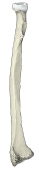 Right radius (anterior)
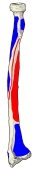 Muscle attachments (anterior)
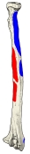 Muscle attachments (posterior)
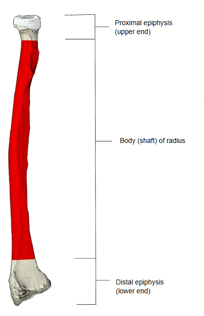
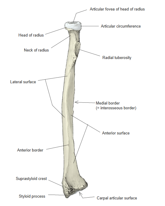
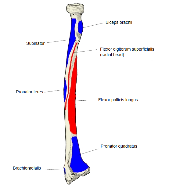
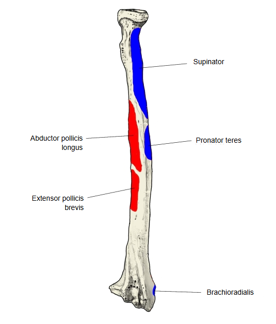
The bowing of the body, convex laterally, carries the lower end of the radius round the ulna when the forearm is turned palm down (pronation); the sharp medial border holds the interosseous membrane that joins the two bones.
Reference websites
[Wikipedia]
· Radius (bone), Wikipedia.
[Websites with illustrations]
· Bones of the left forearm, anterior aspect (Gray's Anatomy, fig. 213) (Wikimedia Commons)

· Bones of the left forearm, posterior surface (Gray's Anatomy, fig. 214) (Wikimedia Commons)

· The radius: proximal, shaft and distal (TeachMeAnatomy)
Where it is found
Related terms
- Proximal epiphysis of radius
- Distal epiphysis of radius
- Radial tuberosity
- Pronator tuberosity
- Anterior border of radius
- Posterior border of radius
- Interosseous border of radius
References and sources
- Further reading: Radius (bone), Wikipedia.
- Federative International Programme for Anatomical Terminology (FIPAT). Terminologia Anatomica, 2nd ed. (TA2), 2019.