Tubercle of trapezium (Latin: tuberculum ossis trapezii)
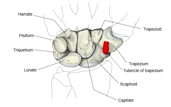
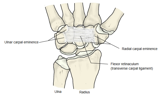
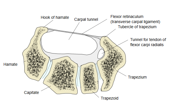
A ridge-like projection on the palmar surface of the trapezium, one of the carpal bones. The flexor retinaculum is attached to it, and it helps to form part of the radial wall of the carpal tunnel.
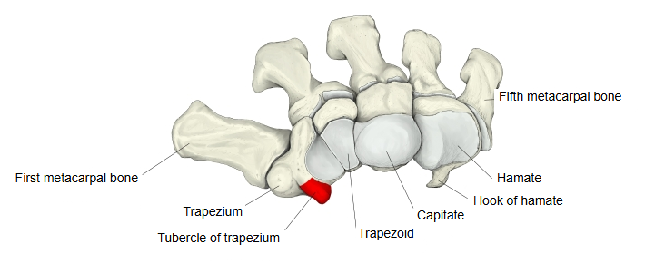
The tubercle stands out at the front of the bone and gives attachment to the superficial part of the flexor retinaculum. Together with the tubercle of the scaphoid it forms the lateral (radial) pillar of the retinaculum; the hook of hamate and the pisiform form the opposite (ulnar) pillar. Along its medial side runs a groove for the tendon of flexor carpi radialis, which is enclosed by a split in the retinaculum. The upper (proximal) end of the bone carries a slightly concave facet for the scaphoid.
It gives partial origin to thenar muscles: opponens pollicis, flexor pollicis brevis and, in some people, abductor pollicis brevis. It can be felt at the base of the thenar eminence, just distal to the tubercle of the scaphoid.
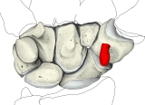 Right carpal bones (palmar)
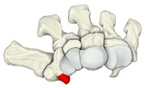 Distal carpal row and metacarpals
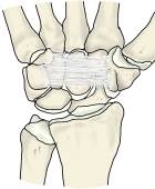 Flexor retinaculum
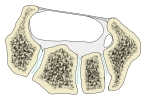 Carpal tunnel (cross-section)
Reference websites
[Wikipedia]
· Trapezium (bone), Wikipedia.
· Carpal tunnel, Wikipedia.
[Websites with illustrations]
· Tubercle of trapezium, left trapezium (palmar view, 3-D render) (Wikimedia Commons)

· Tubercle of trapezium, left trapezium (distal view, 3-D render) (Wikimedia Commons)

· Tubercle of trapezium (animation) (Wikimedia Commons)

Where it is found
Related terms
- Trapezium
- Flexor retinaculum (hand)
- Carpal tunnel
- Hook of hamate
- Carpal bones
- Distal carpal row
- Metacarpal bones
References and sources
- Further reading: Trapezium (bone), Wikipedia.
- Further reading: Carpal tunnel, Wikipedia.
- Federative International Programme for Anatomical Terminology (FIPAT). Terminologia Anatomica, 2nd ed. (TA2), 2019.