Sella turcica (Latin: sella turcica)
Also called Turkish saddle
⇒ A photograph of a Turkish-style horse saddle: Springer (figure)
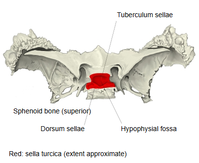
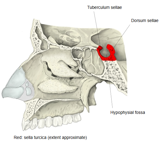
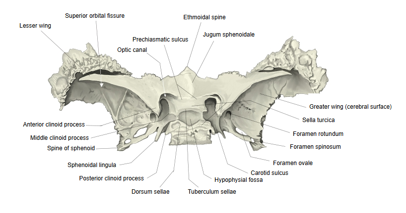
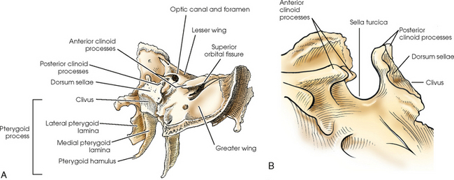
The deepest part, the hypophysial fossa, holds the pituitary gland. In front, the tuberculum sellae has a small middle clinoid process on each side, and in front of it lie the prechiasmatic sulcus and the optic canals. Behind rises a square plate of bone, the dorsum sellae, whose upper corners form the posterior clinoid processes; below and behind, the dorsum continues into the clivus.
The sella turcica is roofed by a fold of dura mater, the diaphragma sellae, and flanked on each side by the cavernous sinus. The sphenoidal sinus lies directly below it, which is why the pituitary gland can be reached through the nose. On a lateral X-ray of the skull its outline is easy to see; enlargement or erosion of it can be a sign of a pituitary tumour.
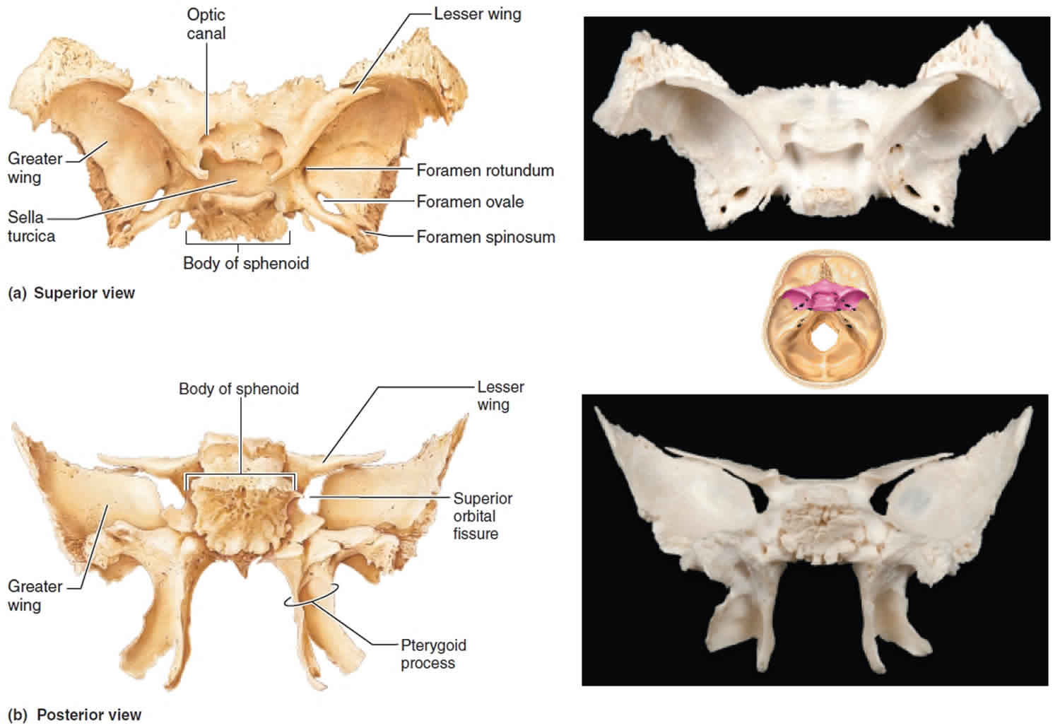
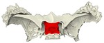 Sphenoid bone (superior)
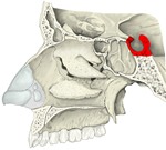 Sagittal section of skull
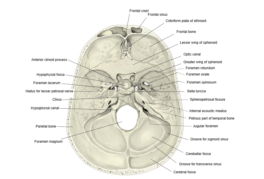
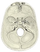 Internal surface of cranial base
 Sphenoid bone (superior), detail

Reference websites
[Wikipedia]
· Sella turcica, Wikipedia.
[Websites with illustrations]
· Sphenoid bone, upper surface (Gray's Anatomy, fig. 145) (Wikimedia Commons)

· Sphenoid bone, superior and posterior views, with the sella turcica (Health Jade)
· Sphenoid bone and the sella turcica with the clinoid processes (Radiology Key)
Where it is found
Related terms
References and sources
- Further reading: Sella turcica, Wikipedia.
- Federative International Programme for Anatomical Terminology (FIPAT). Terminologia Anatomica, 2nd ed. (TA2), 2019.