Articular surface of medial malleolus (Latin: facies articularis malleoli medialis)
Also called the medial malleolar articular surface
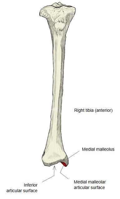
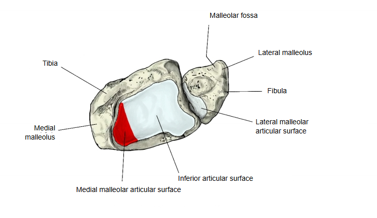
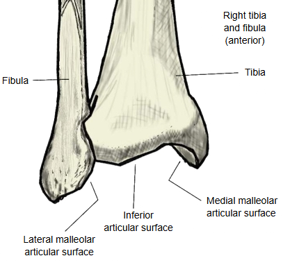
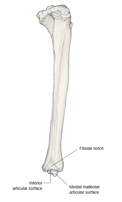
The joint surface on the lateral side of the medial malleolus of the tibia, continuous with the inferior articular surface. It articulates with the trochlea of the talus and forms part of the talocrural (ankle) joint.
It is comma-shaped and meets the inferior articular surface at almost a right angle. It faces the medial malleolar facet on the medial side of the trochlea of the talus. Together with the inferior articular surface and the lateral malleolus of the fibula, it forms the socket (ankle mortise) that grips the talus.
surfaces of
the tibia
- Superior articular surface : the joint surfaces on top of the medial and lateral condyles → knee joint.
- Fibular articular facet : on the back and underside of the lateral condyle → tibiofibular joint.
- Inferior articular surface : on the underside of the lower end → talocrural (ankle) joint.
- Articular surface of medial malleolus : on the lateral side of the medial malleolus, continuous with the inferior articular surface → talocrural (ankle) joint.
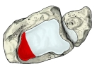 Right tibia and fibula (inferior view)
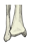 Right tibia and fibula (anterior view)
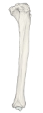 Right tibia (lateral view)
Reference websites
[Wikipedia]
· Malleolus, Wikipedia.
· Ankle, Wikipedia.
[Websites with illustrations]
· Lower end of the tibia, posterior view (photograph) (Wikimedia Commons)

· Tibia: anatomy and clinical notes (Kenhub)
· The tibia: proximal, shaft and distal anatomy (TeachMeAnatomy)
Where it is found
Related terms
- Medial malleolus
- Inferior articular surface of tibia
- Medial malleolar facet
- Talocrural joint
- Superior articular surface of tibia
- Fibular articular facet
- Fibular notch