Cauda helicis (Latin: cauda helicis)
Also called: tail of helix
· It is the tail-like lower end of the helix cartilage, which runs downwards at the back of the auricle towards the lobule, behind and below the antitragus. The antitragohelicine fissure (fissura antitragohelicina) separates it from the antitragus and the lower end of the antihelix. The lobule below it has no cartilage at all. The red area in the figure is approximate: some texts limit the cauda helicis to the pointed tip only; others include more of the lower helix, up to where it merges with the rest of the cartilage.
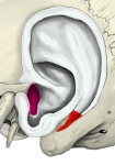 Left auricular cartilage (lateral surface)
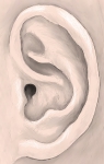 Parts of the auricle
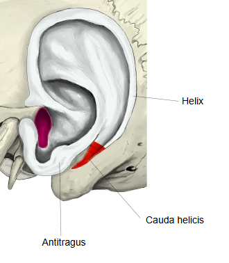
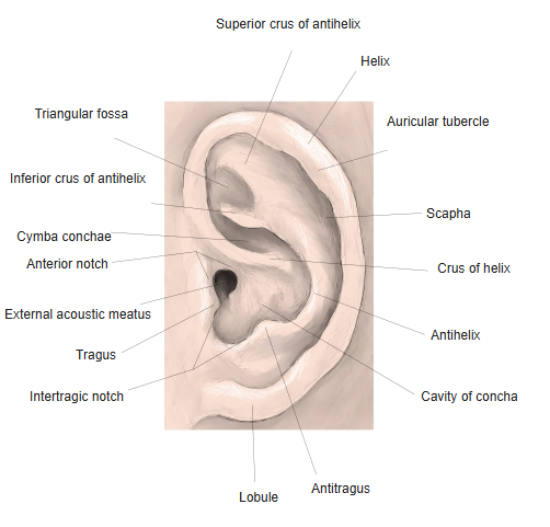
Names of the parts of the auricle
The surface of the auricle is full of ridges and hollows, and its parts have the following names.
Note: the antitragohelicine fissure can also be seen from the lateral surface.
Reference websites
[Wikipedia]
· Helix (ear), Wikipedia.
· Auricle (anatomy), Wikipedia.
[Websites with illustrations]
· Outer surface of the cartilage of the auricle (vintage engraving) (123RF)

· Cartilage of the auricle, medial (cranial) surface, with the cauda helicis (Plastic Surgery Key)

· The auricle, lateral surface (Gray's Anatomy, fig. 904) (Wikimedia Commons)

Related terms
- Helix
- Crus of helix
- Spine of helix
- Tragus
- Auricular cartilage
- Auricle
- Lobule of auricle
- Intertragic notch
- Concha of auricle
- External ear
References and sources
- Further reading: Helix (ear), Wikipedia.
- Further reading: Auricle (anatomy), Wikipedia.
- Federative International Programme for Anatomical Terminology (FIPAT). Terminologia Anatomica, 2nd ed. (TA2), 2019.