Auricular cartilage (Latin: cartilago auriculae)
Also called: cartilage of auricle
The auricular cartilage (elastic cartilage) is the framework of the auricle; it is full of ridges and hollows, and each of them has its own name.
Besides collagen fibres it contains many elastic fibres, so the ear can bend and spring back. The cartilage of the outer part of the external acoustic meatus and of the auditory tube is of the same kind.
Around the auricular cartilage of the auricle lie muscles, ligaments, arteries, veins, lymph nodes and nerves.
· The only part of the auricle without cartilage is the lobule (earlobe) at its lower end; it contains fat and fibrous tissue. The cartilage is continuous with that of the external acoustic meatus. The skin is closely bound to it on the lateral surface and more loosely on the cranial surface.
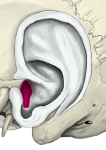 Left auricular cartilage (lateral)
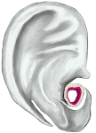 Left auricular cartilage (cranial surface)
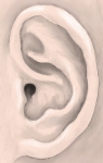 Left auricle (lateral surface)
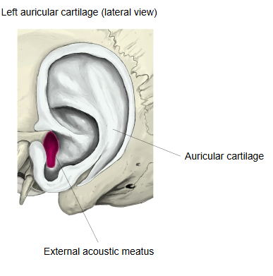
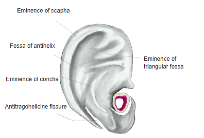
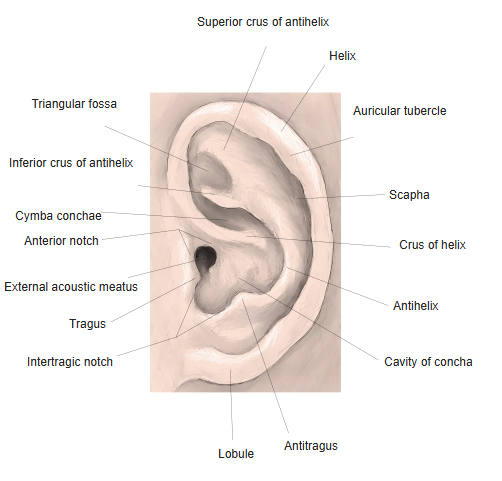
Names of the parts of the auricle
The surface of the auricle is full of ridges and hollows, and its parts have the following names.
Note: the antitragohelicine fissure can also be seen from the lateral surface.
Reference websites
[Wikipedia]
· Auricle (anatomy), Wikipedia.
[Websites with illustrations]
· The auricle, lateral surface (Gray's Anatomy, fig. 904) (Wikimedia Commons)

Related terms
- Tragus
- Lobule of auricle
- Auricular muscles
- Auricular tubercle
- Auricular region
- Concha of auricle
- Auricle
- External ear
- External acoustic meatus
- Extrinsic auricular muscles
- Anterior notch of auricle
- Auricularis posterior
References and sources
- Further reading: Auricle (anatomy), Wikipedia.
- Federative International Programme for Anatomical Terminology (FIPAT). Terminologia Anatomica, 2nd ed. (TA2), 2019.